What is Microspherophakia?
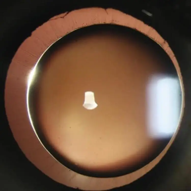
Microspherophakia is a rare abnormality of the crystalline lens, marked by reduced equatorial diameter and increased lens thickness.
People have noticed that when humans are born, their eye lens is almost round, like a ball. It’s about 6.5 mm wide around the middle and 3.5 to 4.0 mm from front to back. But as they grow up, the middle part of the lens gets bigger quickly, and by the time they become adults, it’s about 9 to 10 mm wide.
When they are 20 years old, the front-to-back part of the lens is about 3.7 mm, and when they are 50 years old, it’s about 4.0 mm. This growth makes the lens more round as they get older.
Scientists think that when the fibers that hold the lens are not strong enough, there isn’t enough pull from all sides. This makes the lens stay round instead of becoming a bit curved on both sides. This round shape makes the eyes see things close up blurry. It also makes the space in the front part of the eye shallower, and sometimes, it can lead to a kind of eye problem where the angle in the eye gets blocked. Sometimes, the lens can also move out of place because the fibers holding it are not strong enough.
Read Eye anatomy

![]() The lens is small and spherical in microspherophakia, which may be seen as an isolated familial (dominant) abnormality, or in association with a number of systemic conditions including Marfan and Weill–Marchesani syndromes, hyperlysinemia and congenital rubella.
The lens is small and spherical in microspherophakia, which may be seen as an isolated familial (dominant) abnormality, or in association with a number of systemic conditions including Marfan and Weill–Marchesani syndromes, hyperlysinemia and congenital rubella.
![]() Ocular associations include Peters anomaly and familial ectopia lentis et pupillae.
Ocular associations include Peters anomaly and familial ectopia lentis et pupillae.
![]() Complications can include lenticular myopia, subluxation and dislocation.
Complications can include lenticular myopia, subluxation and dislocation.
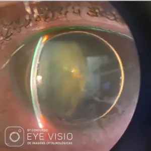
![]() Microphakia is the term used for a lens with a smaller than normal diameter. It may be found in isolation, but may also occur in patients with Lowe syndrome.
Microphakia is the term used for a lens with a smaller than normal diameter. It may be found in isolation, but may also occur in patients with Lowe syndrome.
Credit: Kanski Clinical Ophthalmology.
Microspherophakia cause


Diagnosing Microspherophakia
Ocular examination should include:
- Comprehensive slit-lamp exam: look at lens morphology and angle of the anterior chamber; look for preexisting subluxation
- Fundus exam: check for any glaucomatous damage to the disc
- Peripheral fundus exam: may rule out coexisting retinal pathology
- Intraocular pressure: measure, monitor, and manage appropriately
- Ultrasound biomicroscopy and anterior-segment optical coherence tomography: may help to elucidate biomechanics of angle crowding and angle closure in some patients
- Detailed systemic evaluation: mandatory to rule out syndromic association
- Measure lens thickness and axial length
Clinical Features
Microspherophakia is usually bilateral. The edges of these small-diameter lenses generally can be observed when pupils are fully dilated . Frequently, subluxation is present as well. Other common clinical observations are a high degree of lenticular myopia, defective accommodation, and angle-closure glaucoma.
Most patients who present to an ophthalmologist with microspherophakia will complain of low vision; however, some cases exhibit acute angle-closure glaucoma at initial presentation.
Systemic association of microspherophakia
Other than WMS, microspherophakia has been connected to Alport syndrome, homocystinuria, Klinefelter syndrome, mandibulofacial dysostosis, and Marfan syndrome. It’s also been linked to GEMSS syndrome, which includes glaucoma, ectopia lentis, microspherophakia, stiff joints, and short stature.
Follow us in Facebook
Discover more from An Eye Care Blog
Subscribe to get the latest posts sent to your email.

You must be logged in to post a comment.