External Eye Anatomy
Are you an Eye Professional? Register yourself in our Eye professional directory for Free.
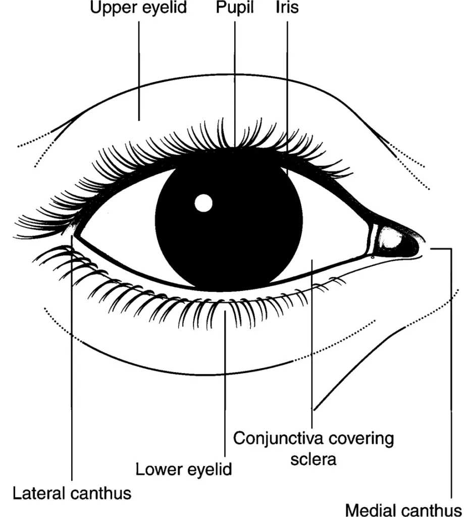
The external parts of the eye work together to protect the eye and all of its internal structures.

external eye anatomy labeled, external eye anatomy diagram
External parts of the eye
The portions of the eye that are normally visible in the palpebral fissures are the cornea and sclera .
Because the cornea is transparent, what is seen on looking at the cornea is the underlying iris and the black opening in the center of the iris is called the pupil .
The sclera forms the white of the eye and is covered by a mucous membrane called the conjunctiva .
The conjunctiva extends from the junction of the cornea and sclera and terminates at the inner portion of the lid margin .
The conjunctiva that covers the eye itself is referred to as the bulbar conjunctiva , whereas the portion that lines the inner surface of the upper and lower lids is called the palpebral conjunctiva .
Eyelids
The upper and lower eyelids form a moist region around the eye, and protect the surface of the eye from injury, infection, and disease.
Eyelashes
The eyelashes are the hairs that grow along the edges of the upper and lower eyelids. The eyelashes protect the eye from foreign particles, such as dust, pollen, and debris. The eyelashes are sensitive to touch and send signals to the eyelids to close when a foreign object comes too close to the eye.
Meibomian glands
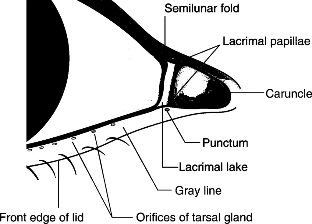
Meibomian glands are the oil glands located inside the eyelids. Their opening pores line the edges of the eyelids, near the eyelashes.
Lacrimal gland
The lacrimal gland creates the water layer of the tears that lubricate, cleanse, nourish, and protect the surface of the eye. This gland is located on the upper lateral wall of the eye, under the eyebrow.
Tear duct
The tear duct, also called the nasolacrimal duct, is located in the inner corner of the eye, and is part of the tear drainage system that goes from the eye, through the back of the nose, and down the throat.
Pupil Size
The pupils are generally equal in size. A normal adult pupil ranges from 2 to 4 millimeters in diameter in bright light. In the darkness, the diameter may range from 4 to 8 millimeters.
Iris
The iris in humans is the colored (typically brown, blue, or green) area, with the pupil (the circular black spot) in its center, and surrounded by the white sclera.(wiki)
Sclera
The sclera, or white of the eye, is a protective covering that wraps over most of the eyeball. It extends from the cornea in the front to the optic nerve in the back.
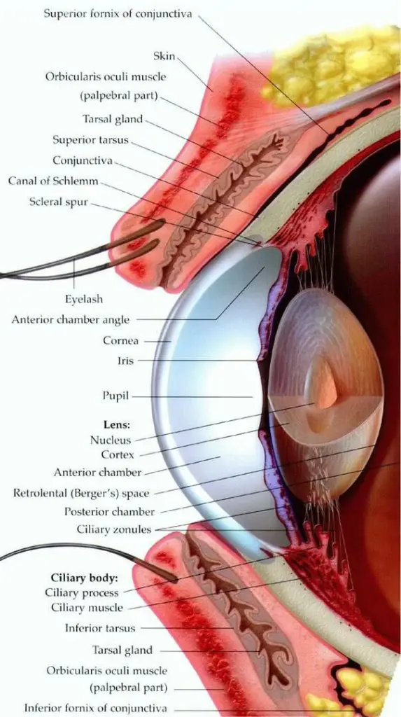
A detailed diagram of inner eye

external eye anatomy accessory structures
Here is a detailed info on External Eye Anatomy
In this diagram we will be able to locate the external Eye parts with reference to Eye’s cross section diagram.
Here you will be able to locate the following Parts:
- Superior fornix Of conjunctiva
- Skin
- Orbicularis oculi muscle (palpebral part)
- Tarsal gland
- Superior tarsus
- Conjunctiva
- Canal of Schlemm
- Scleral spur
- Eyelash
- Anterior chamber angle
- Cornea
- Iris
- pupil
- Nucleus
- Cortex
- Anterior chamber
- Retrolental (Berger’s) space
- Posterior chamber
- Ciliary zonules
- Ciliary body:
- Ciliary process
- Ciliary muscle
- Inferior tarsus
- Tarsal gland
- Orbicularis oculi muscle (palpebral part)
- Inferior fornix Of conjunctiva

- The orbicularis oculi muscle is a ringlike band of muscle, called a sphincter muscle, that surrounds the eye. It lies in the tissue of the eyelid and causes the eye to close or blink.
- Meibomian glands (also called tarsal glands, palpebral glands, and tarsoconjunctival glands) are sebaceous glands along the rims of the eyelid inside the tarsal plate. They produce meibum, an oily substance that prevents evaporation of the eye’s tear film.
- The anterior chamber is the space between the cornea and the iris. It is filled with aqueous humor, a water fluid produced by the ciliary body that provides nutrients and oxygen for the lens and cornea.
- The posterior chamber is the space between the posterior surface of the iris and the anterior surface of the lens. It is filled with aqueous humor.
- The vitreous chamber is the space between the posterior surface of the lens and the retina. It is filled with the vitreous body, a transparent gel that is made of 99% water, in addition to extremely delicate type II collagen fibers, hyaluronic acid, and electrolytes.
- Fornix inferior conjunctivae : The line of reflection of the conjunctiva from the lower eyelid is the inferior conjunctival fornix, it forms the junction between the bulbar and palpebral conjunctivas. It is loose and flexible, allowing the free movement of the lids and eyeball.
- Berger’s space is an anatomical but normally invisible space behind the crystalline lens. It is formed by the posterior lens surface and the anterior vitreous face
- Ciliary zonules are a ring of fibrous structures anchoring the ciliary body with the lens of the eye. These are the structures that help to maintain the position of the lens in the optical path, and anchor muscles that change the shape of the lens to alter focus.
- The ciliary body is a ring-shaped thickening of tissue inside the eye that divides the posterior chamber from the vitreous body.
- The ciliary processes are formed by the inward folding of the various layers of the choroid
- The ciliary muscle is an intrinsic muscle of the eye formed as a ring of smooth muscle in the eye’s middle layer (vascular layer). It controls accommodation for viewing objects at varying distances and regulates the flow of aqueous humor into Schlemm’s canal.
- The tarsi ( tarsal plates) are two thin, elongated plates of dense connective tissue, about 2.5 cm. in length; one is placed in each eyelid, and contributes to its form and support
Discover more from An Eye Care Blog
Subscribe to get the latest posts sent to your email.

You must be logged in to post a comment.