Surfaces and layers of the iris:

iris ciliary body
What does the iris do?
The iris is the colored part of the eye that lies between the cornea and the lens. It is responsible for regulating the amount of light that enters the eye by controlling the size of the pupil. The surface of the iris is smooth and has a slightly convex shape.
Surfaces and layers of the iris:
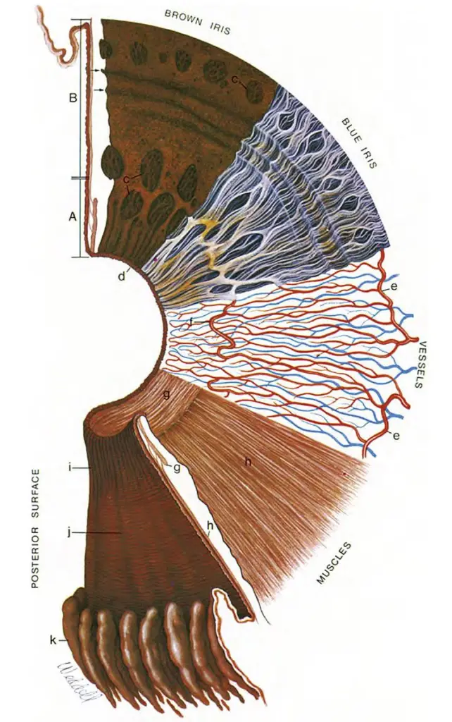
![]() Beginning at the upper left and proceeding clockwise, the iris cross-section shows the pupillary (A) and ciliary portions (B), and the surface view shows a brown iris with its dense, matted anterior border layer.
Beginning at the upper left and proceeding clockwise, the iris cross-section shows the pupillary (A) and ciliary portions (B), and the surface view shows a brown iris with its dense, matted anterior border layer.
![]() Circular contraction furrows are shown (arrows) in the ciliary portion of the iris. Fuchs’ crypts (c) are seen at either side of the collarette in the pupillary and ciliary portion and peripherally near the iris root. The pigment ruff is seen at the pupillary edge (d).
Circular contraction furrows are shown (arrows) in the ciliary portion of the iris. Fuchs’ crypts (c) are seen at either side of the collarette in the pupillary and ciliary portion and peripherally near the iris root. The pigment ruff is seen at the pupillary edge (d).
![]() The blue iris surface shows a less dense anterior border layer and more prominent trabeculae.
The blue iris surface shows a less dense anterior border layer and more prominent trabeculae.
![]() The iris vessels are shown beginning at the major arterial circle in the ciliary body (e).
The iris vessels are shown beginning at the major arterial circle in the ciliary body (e).
![]() Radial branches of the arteries and veins extend toward the pupillary region. The arteries form the incomplete minor arterial circle (f), from which branches extend toward the pupil, forming capillary arcades.
Radial branches of the arteries and veins extend toward the pupillary region. The arteries form the incomplete minor arterial circle (f), from which branches extend toward the pupil, forming capillary arcades.
![]() The sector below it demonstrates the circular arrangement of the sphincter muscle (g) and the radial processes of the dilator muscle (h).
The sector below it demonstrates the circular arrangement of the sphincter muscle (g) and the radial processes of the dilator muscle (h).
![]() The posterior surface of the iris shows the radial contraction furrows (i) and the structural folds of Schwalbe (j).
The posterior surface of the iris shows the radial contraction furrows (i) and the structural folds of Schwalbe (j).
![]() Circular contraction folds also are present in the ciliary portion. The pars plicata of the ciliary body is at k.
Circular contraction folds also are present in the ciliary portion. The pars plicata of the ciliary body is at k.
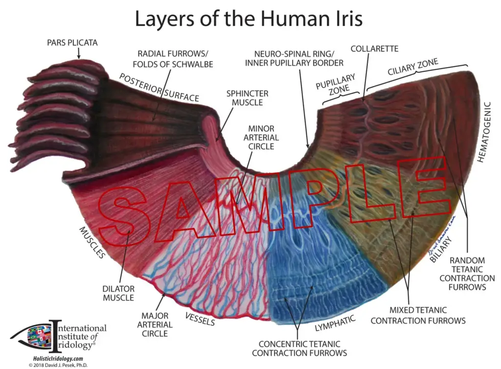
The iris is composed of several layers, each with its own distinct properties and functions. Starting from the outermost layer and moving inward, these layers are:
- Anterior border layer: This is the outermost layer of the iris and is composed of a thin layer of cells that form a ring around the iris. These cells help to anchor the iris to the surrounding tissue.
- Stroma: This is the thickest layer of the iris and is composed of connective tissue and pigmented cells. The pigmented cells give the iris its color and help to absorb excess light that enters the eye. The connective tissue provides structural support for the iris.
- Posterior border layer: This is a thin layer of cells that lines the back of the stroma. It helps to separate the iris from the ciliary body, which produces the aqueous humor that fills the front of the eye.
- Iris sphincter muscle: This is a circular muscle that surrounds the pupil and helps to control its size. When this muscle contracts, the pupil becomes smaller, and when it relaxes, the pupil becomes larger.
- Iris dilator muscle: This is a radial muscle that lies beneath the iris sphincter muscle. It helps to dilate the pupil by pulling the iris outward.
- Anterior surface: This is the innermost surface of the iris and faces the front of the eye. It is covered by a layer of cells called the iris epithelium, which helps to maintain the shape of the iris and protect its delicate internal structures.
Together, these layers work in concert to regulate the amount of light that enters the eye and to maintain the shape and function of the iris. Understanding the anatomy and function of the iris is essential for diagnosing and treating a wide range of eye conditions and diseases.

See: Anatomy of iris and ciliary body
Credit: From Hogan MJ, Alvarado JA, Weddell JE: Histology of the human eye, Philadelphia, 1971, Saunders.
Follow us at Facebook.com
Discover more from An Eye Care Blog
Subscribe to get the latest posts sent to your email.


You must be logged in to post a comment.