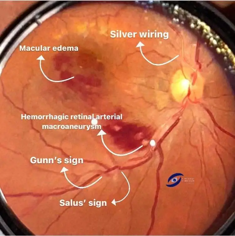
Gunn’s sign and salus sign
Understanding Hypertensive Retinopathy and its Effects on the Optic Nerve
Hypertensive retinopathy is a condition characterized by retinal changes due to chronic high blood pressure. It holds particular importance due to its potential to harm the optic nerve, cause retinal deflection, and affect arteriovenous crossings. Two crucial clinical signs associated with hypertensive retinopathy are Gunn’s sign and Salus sign.
What Is Hypertensive Retinopathy?
Hypertensive retinopathy results from persistent high blood pressure, where the force of blood against the blood vessel walls remains consistently high. This can have detrimental effects on the blood vessels in the retina, the light-sensitive layer at the back of the eye.
The Impact on the Optic Nerve
Uncontrolled high blood pressure can lead to damage in the optic nerve. The optic nerve plays a vital role in vision, transmitting visual information from the eye to the brain. When hypertension affects the optic nerve, it can result in vision problems and potentially even vision loss.
Gunn’s sign and salus sign in retinal image
Gunn’s sign and Salus sign are two important signs that can be observed in retinal images. These signs indicate the presence of certain pathological conditions in the eye.

Gunn’s Sign & Salus’s Sign at retinal arteriovenous crossings in Hypertensive Retinopathy image . Photo credit: visus.formacion.
diagnosis:
- Gunn’s sign: Tapering of veins on either side of arteriovenous crossing.
- Bonnet sign: Dilatation of the veins distal to the arteriovenous crossing.
- Salus sign: Deflection of veins at arteriovenous crossing.
Gunn’s sign and salus sign in Hypertensive retinopathy.

In hypertensive retinopathy, there is an increased risk of retinal deflection, where the retina is pushed out of its normal position. This disruption can affect the proper functioning of the retina, contributing to vision issues.
Arteriovenous crossings, or points where arteries and veins in the retina intersect, can become distorted and compromised due to increased pressure in the blood vessels. This can further exacerbate retinal damage and impact overall eye health.
Both Gunn’s sign and Salus sign are indicators of hypertensive retinopathy, reflecting damage to the retinal arterioles caused by high blood pressure. Gunn’s sign typically appears later in severe hypertension cases with end-organ damage, while Salus sign emerges earlier in mild to moderate hypertension. Ophthalmoscopy reveals both signs, making them important diagnostic and monitoring tools for ophthalmologists and optometrists managing patients with hypertensive retinopathy.

Gunn’s sign:
Gunn’s sign pertains to the narrowing of retinal arterioles observed in hypertensive retinopathy. The narrowing can be so severe that the arterioles appear as “silver wires” on ophthalmoscopy. This narrowing results from thickened arteriolar walls due to increased blood pressure, reducing the lumen size and enhancing reflectivity. Gunn’s sign, also known as the “silver-wire” arteriole sign, is a late sign of hypertensive retinopathy associated with severe hypertension and organ damage.
Thickened arteriole walls and sclerosis are believed to compress the vein at crossing points, obstructing blood flow and causing vein tapering at the crossing, commonly known as Gunn’s sign. It’s identifiable by examining retinal arterioles for increased reflectivity along their walls.
excrept from Kanski JJ, Bowling B. Clinical Ophthalmology: A Systematic Approach. Edinburgh: Elsevier; 2016. Chapter 19: Hypertensive Retinopathy, pp. 263-268, says-
Gunn’s sign refers to the narrowing of the retinal arterioles seen in hypertensive retinopathy, which can be so severe that the arterioles appear to be “silver wires” on ophthalmoscopy. This sign is caused by the thickening of the arteriolar walls due to the increased pressure of blood flow, which reduces the lumen size and makes the walls more reflective. Gunn’s sign is considered a late sign of hypertensive retinopathy and is associated with severe hypertension and end-organ damage.
Gunn’s sign, also known as the “silver-wire” arteriole sign, is a sign of hypertensive retinopathy, which is a manifestation of high blood pressure.
Thickening of the arteriole wall and/or sclerosis are thought to compress the vein at a crossing point, impeding blood flow, causing a tapering of the vein at the crossing. This tapering is commonly referred to as Gunn’s sign.
In this sign, the walls of the retinal arterioles appear to be more reflective and silver-like than normal, due to thickening of the arteriolar walls. To identify Gunn’s sign, one needs to examine the retinal arterioles in the retinal image and look for any regions of increased reflectivity along the arteriolar walls.
Salus sign:
Salus sign refers to the interruption or splitting of the arteriolar light reflex observed in hypertensive retinopathy, which can give the arterioles a “copper-wire” appearance on ophthalmoscopy. Thickened arteriolar walls and irregular surface due to increased blood pressure cause this sign. Salus sign, also known as the “copper-wire” arteriole sign, is an early indicator of hypertensive retinopathy linked to mild to moderate hypertension.
Pressure from the artery can displace the vein at the crossing, causing it to enter and exit the crossing at a 90° angle, a phenomenon referred to as Salus’s sign. To identify Salus sign, you need to examine the retinal arterioles for regions of increased opacity and a copper-like appearance along the arteriolar walls.
Excrept from Kanski JJ, Bowling B. Clinical Ophthalmology: A Systematic Approach. Edinburgh: Elsevier; 2016. Chapter 19: Hypertensive Retinopathy, pp. 263-268, says
Salus sign refers to the splitting of the arteriolar light reflex seen in hypertensive retinopathy, which can give the arterioles a “copper-wire” appearance on ophthalmoscopy. This sign is caused by the thickening of the arteriolar walls and the irregularity of the surface due to the increased pressure of blood flow.
The light reflex is interrupted or broken where the wall is thickened, giving the appearance of a wire with a copper-colored coating. Salus sign is considered an early sign of hypertensive retinopathy and is associated with mild to moderate hypertension.
Salus sign, also known as the “copper-wire” arteriole sign, is another sign of hypertensive retinopathy. In this sign, the walls of the retinal arterioles appear to be more opaque and copper-like than normal, due to thickening and sclerosis of the arteriolar walls.
The pressure from the artery can also displace the vein at the crossing, causing the vein to enter and exit the crossing at a 90° angle. This phenomena is commonly referred to as Salus’s sign.
To identify Salus sign, one needs to examine the retinal arterioles in the retinal image and look for any regions of increased opacity and copper-like coloration along the arteriolar walls.

Salus sign as markers of increased intracranial pressure in hypertensive retinopathy
Managing hypertensive retinopathy based on Gunn’s and Salus sign findings:
As primary eye care physicians, optometrists may be the first to see hypertensive changes in patients who are undiagnosed with this condition. Routinely checking blood pressure in the optometric practice will assist the clinician to make an appropriate diagnosis and referral in a timely manner.
Understanding Hypertensive Retinal changes
Watch video: https://fb.watch/nTyKfqm2YH/
Conclusion
Hypertensive retinopathy is a condition resulting from high blood pressure, with far-reaching consequences for eye health. It can harm the optic nerve, cause retinal deflection, and affect arteriovenous crossings. Monitoring and managing blood pressure is crucial in preventing and mitigating the effects of hypertensive retinopathy on the eyes.
If you have high blood pressure, it’s vital to work with a healthcare professional to keep it under control and protect your eye health. Optometrists can play a key role by routinely checking blood pressure to diagnose and refer patients with hypertensive retinopathy promptly.
Follow us in Facebook for more updates

