Cones, rods in the retina may still retain visual function
Cone photoreceptors in retinal degeneration have been thought to be dormant. However, new research suggests cone photoreceptors in a degenerating retina may continue producing responses to light. Researchers recorded downstream signals from the retina that indicate visual processing was not as compromised as might be expected.

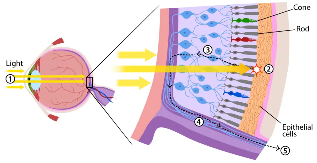
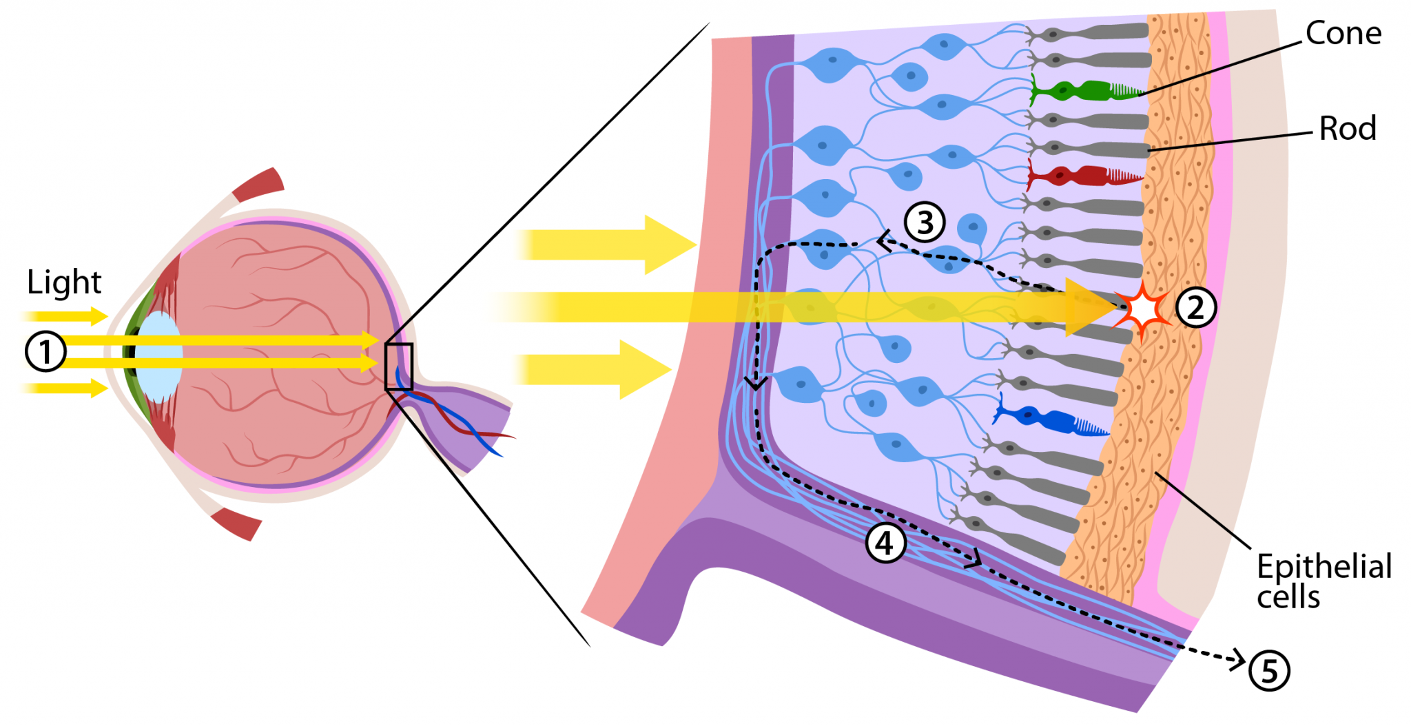
In the retina, the cells responsible for the visual experience are rods and cones.These cells are called photoreceptors and they absorb and convert light into electric signals.Rods are active in dim light. Cones are active in daylight and help a person see colors.
Alapakkam Sampath, the chair of ophthalmology at the the University of California Los Angeles’ Jules Stein Eye Institute and a professor at the David Geffen School of Medicine at UCLA, has spent the past quarter of a century studying how photoreceptors in the retina work.
Initially, retinitis pigmentosa affects the rods, which causes night vision issues. As the rods die off, the ailment begins to affect the cones, leading to blindness.
“Typically in the literature they’ve always been called dormant cones,” Sampath explained. “The dormancy has embedded in it the notion that they’re not doing anything.”
Sampath and the other researchers set out to understand the “nature of the dysfunction.”
“Because I think this is the way to figure out whether [the photoreceptors] can be repaired or to what extent they could be rescued,” he said.
To do this, the researchers “studied the photoreceptors that are left when other photoreceptors are degenerating,” Sampath said.
Specifically, they made patch clamp recordings from cells in the central region of the retina in rd10miceTrusted Source, which model autosomal recessive retinitis pigmentosa. The rods on the mice cell had mostly died and the cones had lost their outer segments and pedicles.
The patch clamp method is a refined electrophysiological technique that measures the membrane potential as well as the amount of current passing across the cell membrane.
Additionally, researchers used multi-electrode arraysTrusted Source to make recordings of retinal responses to presented visual stimuli.
The researchers said they were surprised to find many of the cones were able to respond to light.
“We showed that they were remarkably still active, although a lot less sensitive than normal,” Sampath said.
Researchers observed light response in four out of four cones in mice that were 3.5 weeks old as well as 7 of 10 cones in mice that were 6 weeks old and 1 out of 3 cones at 9 weeks old.
The sensitivity of the cones was about 100-fold to 1,000-fold less than normal.
The cells also displayed many of the features of normal cones. This included similar resting membrane potentialTrusted Source, which refers to the electrical potential difference across the plasma membrane when the cell is at rest and a normal synaptic Ca2+ current.
Compensatory mechanism?
Greg Field, an adjunct associate professor of neurobiology at the Duke School of Medicine in North Carolina, led a part of the study that looked at retinal ganglion cellsTrusted Source, which are responsible for projecting visual stimuli to the brain.
“What Greg’s laboratory worked on is recording signals from all of the residual cones as they are represented in the ganglion cells,” Sampath explained. “What he found was surprisingly that the loss of sensitivity at the level of the ganglion cells was not as much as you might predict based on the loss of sensitivity we saw in the photoreceptors, so there must be some type of adaptation or compensatory mechanism that’s trying to protect signals… The brain and the retina as an extension of the brain tries super hard to ensure that function is protected for as long as possible at the highest quality possible.”
These cones might be key to figuring out how to repair lost sight.
“What Greg showed in his study is that the spatial properties of the cells as well as the time scale on which they’re active are not changed that much,” Sampath said. “It’s only that the sensitivity is reduced so the potential is there is if the sensitivity of the cones could be restored or increased… you might actually have nearly normal daytime vision. So these residual cones may be a great conduit for rescuing visual experience that must be debilitating. We don’t know how how much more visual loss there is. We haven’t done the behavioral tests on these mice under these conditions, but the presumption is that… their vision is far less sensitive.”
“So the significant science of the article is that even after nerve cells, in this case retinal photoreceptors, have lost their ability to transmit a detectable signal by the organism, even after the [rodent] is blind, this study is demonstrating that there still is light responsiveness of the photoreceptor. And therefore, it speculates that there should be some way of amplifying that responsiveness or interrupting the degenerative process to potentially restore vision.”
The study was published in medicalnewstoday
Discover more from An Eye Care Blog
Subscribe to get the latest posts sent to your email.


You must be logged in to post a comment.