Candle Wax Dripping Lesions” in ocular sarcoidosis
Candle wax dripping lesion in ocular sarcoidosis is a rare manifestation of this systemic disease. Sarcoidosis is a multisystem inflammatory disorder that can affect any organ, including the eyes. The diagnosis of ocular sarcoidosis can be challenging, and the candle wax dripping lesion is just one of the many clinical signs that can help ophthalmologists make a diagnosis.
See Other Eye Diseases
‘‘Candle Wax Dripping’’ Lesions in Sarcoidosis:
Sarcoidosis is a systemic inflammatory disease characterized by the formation of noncaseating granulomas in affected organs, most commonly the lungs, lymph nodes, skin, and eyes
Ocular involvement in sarcoidosis is present in up to 80 percent of patients and is frequently manifested before diagnosis of the underlying systemic disease
According to the book “Uveitis: A Practical Guide to the Diagnosis and Treatment of Intraocular Inflammation” by Robert B. Nussenblatt and Scott M. Whitcup, the candle wax dripping lesion is characterized by the accumulation of white-grayish material on the inferior cornea that resembles dripping candle wax. This material is composed of aggregates of inflammatory cells and proteins.
The candle wax dripping lesion is not pathognomonic of ocular sarcoidosis, as it can also be seen in other inflammatory conditions such as tuberculosis and fungal infections. However, it is a highly suggestive sign of ocular sarcoidosis when combined with other clinical signs such as anterior uveitis, mutton-fat keratic precipitates, iris nodules, and vitritis.
The diagnosis of ocular sarcoidosis requires a thorough evaluation that includes a detailed medical history, physical examination, laboratory tests, and imaging studies. According to the book “Clinical Ophthalmic Echography: A Case Study Approach” by Roger P. Harrie, imaging studies such as ultrasound biomicroscopy and anterior segment optical coherence tomography can be useful in detecting the candle wax dripping lesion.
The treatment of ocular sarcoidosis depends on the severity of the disease and the organs involved. According to the book “Uveitis and Immunological Disorders” by Sunil K. Srivastava and Phuc LeHoang, topical and systemic corticosteroids are the mainstay of treatment for ocular sarcoidosis. Immunosuppressive agents such as methotrexate, azathioprine, and mycophenolate mofetil may be added in severe cases.
In conclusion, the candle wax dripping lesion is a rare but highly suggestive sign of ocular sarcoidosis. Ophthalmologists should be aware of this clinical sign and combine it with other clinical and imaging findings to make a diagnosis. A multidisciplinary approach involving ophthalmologists, pulmonologists, and rheumatologists is essential for the management of this complex disease.
Candle Wax Dripping’’ Lesions in Sarcoidosis:

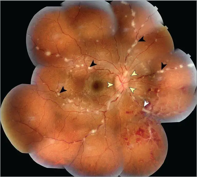
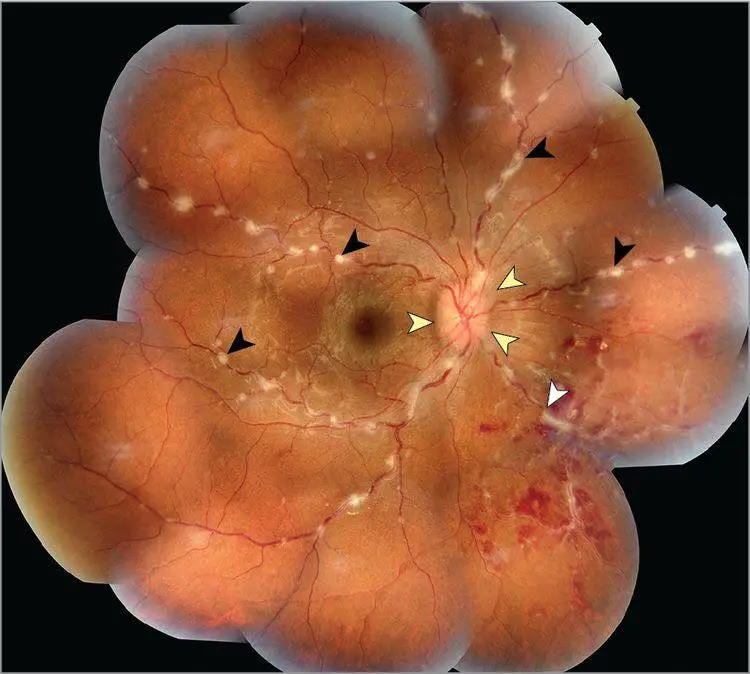
presence of nodular and/or segmental periphlebitis, also known as “candle wax dripping”
![]() A woman in her late 20s presented with a 2-week history of blurred vision in both eyes.
A woman in her late 20s presented with a 2-week history of blurred vision in both eyes.![]() Ophthalmoscopy results revealed bilateral optic disc edema and multifocal and segmented periphlebitis with the characteristic “candle wax dripping,” especially in the right eye, causing a branch vein occlusion.
Ophthalmoscopy results revealed bilateral optic disc edema and multifocal and segmented periphlebitis with the characteristic “candle wax dripping,” especially in the right eye, causing a branch vein occlusion.![]() “Candle wax dripping” lesions are typically seen during acute ocular sarcoidosis, and this finding is usually associated with a poor long-term prognosis with more frequent relapses.
“Candle wax dripping” lesions are typically seen during acute ocular sarcoidosis, and this finding is usually associated with a poor long-term prognosis with more frequent relapses.
Credit pic : Lezrek et al., JAMA Ophthalmol. 2017;135(8):e171845.
References:
- Nussenblatt RB, Whitcup SM. Uveitis: A Practical Guide to the Diagnosis and Treatment of Intraocular Inflammation. New York: Springer; 2012.
- Harrie RP. Clinical Ophthalmic Echography: A Case Study Approach. New York: Springer; 2009.
- Srivastava SK, LeHoang P. Uveitis and Immunological Disorders. New York: Springer; 2014.
Follow us in Facebook for more updates
Discover more from An Eye Care Blog
Subscribe to get the latest posts sent to your email.


You must be logged in to post a comment.