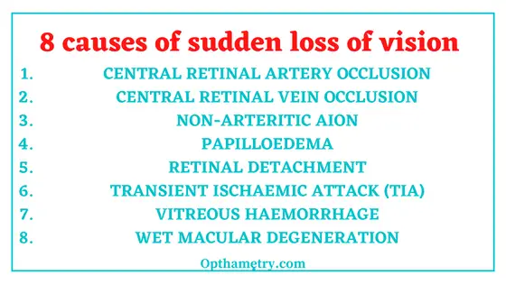
8 causes of sudden loss of vision

8 causes of sudden loss of vision are as follows:
- CENTRAL RETINAL ARTERY OCCLUSION
- CENTRAL RETINAL VEIN OCCLUSION
- NON-ARTERITIC AION
- PAPILLOEDEMA
- RETINAL DETACHMENT
- TRANSIENT ISCHAEMIC ATTACK (TIA)
- VITREOUS HAEMORRHAGE
- WET MACULAR DEGENERATION
Let us read the causes of loss of vision in detail:
See Eye anatomy

central retinal artery occlusion
When one of the vessels that carry blood to your eye’s retina gets blocked, it can cause you to lose your eyesight. This problem often happens suddenly and without any pain. This is called a central retinal artery occlusion (CRAO).
Who is at risk for central retinal artery occlusion?
High blood pressure and aging are the main risks for CRAO. Glaucoma and diabetes can also raise your risk, as can problems in which your blood is thicker and stickier than normal. In women, the problem has been linked to the use of birth control pills.
2. Central Retinal Vein Occlusion (CRVO)
CRVO happens when a blood clot blocks the flow of blood through the retina’s main vein.
Who Is at Risk for CRVO?
CRVO usually happens in people who are aged 50 and older.
People who have the following health problems have a greater risk of CRVO:
high blood pressure
diabetes
glaucoma
hardening of the arteries (called arteriosclerosis)
3. non-arteritic anterior ischemic optic neuropathy
Non-arteritic anterior ischemic optic neuropathy (NAION or NA-AION) is caused by decreased blood flow to the front part of the optic nerve. It causes optic nerve swelling and sudden vision loss. NAION typically affects one eye, although the other eye sometimes suffers similar loss months or years later (about a 15% risk of second eye involvement within 5 years). If there is sudden severe blood loss or severe and prolonged drop in blood pressure, both eyes may be affected together.
it happens more often in patients born with small optic discs (the front part of the optic nerve that can be seen within the eye). This crowded optic disc structure (often referred to as a small cup-to-disc ratio or a “disc at risk”) makes the nerve more vulnerable to blood supply problems.
4. Papilledema
Papilledema is when pressure in your brain makes your optic nerve swell.
If your doctor believes a brain condition is causing papilledema, they’ll do additional tests. Your doctor may order an MRI test or a CT scan of your head to check for tumors or other abnormalities in your brain and skull.
5. Retinal detachment
Retinal detachment separates the retinal cells from the layer of blood vessels that provides oxygen and nourishment to the eye. The longer retinal detachment goes untreated, the greater your risk of permanent vision loss in the affected eye.
Warning signs of retinal detachment may include one or all of the following: reduced vision and the sudden appearance of floaters and flashes of light. Contacting an eye specialist (ophthalmologist) right away can help save your vision.
Retinal detachment itself is painless. But warning signs almost always appear before it occurs or has advanced, such as:
- The sudden appearance of many floaters — tiny specks that seem to drift through your field of vision
- Flashes of light in one or both eyes (photopsia)
- Blurred vision
- Gradually reduced side (peripheral) vision
- A curtain-like shadow over your field of vision
6. Transient ischemic attacks
Transient ischemic attacks usually last a few minutes. Most signs and symptoms disappear within an hour, though rarely symptoms may last up to 24 hours. The signs and symptoms of a TIA resemble those found early in a stroke and may include sudden onset of:
Weakness, numbness or paralysis in the face, arm or leg, typically on one side of the body
Slurred or garbled speech or difficulty understanding others Blindness in one or both eyes or double vision ,Vertigo or loss of balance or coordination
7. vitreous hemorrhage
A vitreous hemorrhage occurs when blood enters the vitreous of the eye, which is the thick, clear fluid that fills the eye and helps maintain its shape. A vitreous hemorrhage may affect your vision if it prevents light from reaching the retina, a light-sensitive tissue in the back of the eye responsible for eyesight
Vitreous hemorrhage can appear in many different ways. Symptoms can include unilateral floaters and/or vision loss. Some people with a mild case experience certain symptoms earlier on. These signs include:
- Floaters
- Haze
- Cobwebs
- A red hue
- Shadows
The symptoms may be worse in the morning due to blood pooling in your eye while lying down. Vitreous hemorrhage symptoms are not usually painful, and they may happen suddenly.(ref 2)
8. Wet macular degeneration
Wet macular degeneration is a long-lasting eye disorder that causes blurred vision or a blind spot in the central vision. It’s usually caused by blood vessels that leak fluid or blood into the macula.
Wet macular degeneration is one of two types of age-related macular degeneration. The other type, dry macular degeneration, is more common and less severe. The wet type always begins as the dry type.
Early detection and treatment of wet macular degeneration may help reduce vision loss. In some instances, early treatment may recover vision.
So these were the 8 causes of sudden loss of vision. If you have any of these symptoms, you must check with your doctor at earliest.
References
Discover more from An Eye Care Blog
Subscribe to get the latest posts sent to your email.

You must be logged in to post a comment.