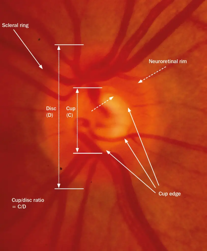
Optic disc and neuro retinal rim photographs
The optic disc is responsible for transmitting visual information from the retina to the brain. It is also known as the optic nerve head and is located in the back of the eye, where the optic nerve exits the eye and connects to the brain. The neuro retinal rim is the outer edge of the optic disc, which contains the nerve fibers that make up the optic nerve.
The neuro retinal rim is an important component of the optic disc, as it provides support and protection to the optic nerve fibers. It is composed of a thin layer of tissue that surrounds the optic disc and is made up of a combination of glial cells and connective tissue.

Neuro retinal rim Thickness
In clinical practice, Neuro retinal rim Thickness is measured using various imaging techniques such as optical coherence tomography (OCT) and scanning laser ophthalmoscopy (SLO). These techniques allow ophthalmologists to obtain high-resolution images of the optic nerve head and accurately measure the thickness of the neuro retinal rim.
A healthy neuro retinal rim is typically at least 0.1 mm thick, although the actual thickness can vary depending on factors such as age and race. Thinning of the neuro retinal rim is a common sign of glaucoma, a group of eye diseases that can cause damage to the optic nerve and lead to vision loss.
In addition to glaucoma, other conditions that can affect the thickness of the neuro retinal rim include optic neuritis, multiple sclerosis, and traumatic optic neuropathy. These conditions can cause inflammation or damage to the optic nerve, leading to a loss of axons and thinning of the neuro retinal rim.
Rods and cones in the retina may still retain visual function despite eyesight loss
Role of neuro retinal rim
- Neuro retinal rim is to help maintain the structural integrity of the optic disc. This is important because any damage to the optic disc can result in vision loss or even blindness. The neuro retinal rim helps to protect the optic nerve fibers by providing a cushion against external pressures and forces.
- Neuro retinal rim helps to help regulate the flow of blood and nutrients to the optic nerve fibers. The blood vessels that supply the optic nerve fibers with oxygen and nutrients are located within the neuro retinal rim. These vessels are critical for the health and survival of the optic nerve fibers, and any disruption to their blood supply can lead to vision loss.
Disease due to abnormal neuro retinal rim
Several different diseases and conditions can affect the neuro retinal rim and the optic disc. One of the most common is glaucoma, which is a group of eye diseases that damage the optic nerve and can lead to vision loss and blindness. In glaucoma, the pressure inside the eye increases, which can cause damage to the optic nerve fibers and the neuro retinal rim. Other conditions that can affect the optic disc and neuro retinal rim include optic neuritis, optic atrophy, and papilledema.
Let us see some Optic disc and neuro retinal rim photographs
The cup disc ratio in Retina
Cup disc ratio is an important parameter used in ophthalmology to assess the health of the optic nerve. It is the ratio between the size of the cup, which is the depression in the center of the optic disc, and the size of the optic disc itself. A larger cup disc ratio indicates a higher risk of glaucoma and other optic nerve related diseases.
n the book “Ophthalmology” by Myron Yanoff and Jay S. Duker, the authors describe the cup disc ratio as a key factor in diagnosing and monitoring glaucoma. They note that a cup disc ratio of 0.3 or less is considered normal, while a ratio of 0.4 or higher is indicative of glaucoma. However, they caution that the interpretation of the cup disc ratio should be done in conjunction with other clinical findings and tests.


The scleral rim

The scleral rim, also called the limbus, is the border between the cornea and the sclera of the eye. It is a transitional zone where the transparent cornea meets the opaque white sclera. The scleral rim plays an important role in the structural integrity of the eye and is also a critical location for certain surgical procedures.
One of the primary functions of the scleral rim is to anchor the muscles that control eye movement. The six extraocular muscles that move the eye are attached to the scleral rim, giving them a stable point of origin. This allows the muscles to exert precise control over the position and movement of the eye.
The scleral rim is an important location for certain surgical procedures, such as corneal transplantation and glaucoma surgery. During these procedures, the surgeon must carefully manipulate the scleral rim to access the underlying structures of the eye. The scleral rim is also a common site for the placement of scleral contact lenses, which can be used to correct certain refractive errors.
The scleral rim is also a source of stem cells that can be used to regenerate damaged or diseased corneal tissue. These stem cells are located in a specialized niche at the limbus and have the ability to differentiate into various cell types, including corneal epithelial cells, which are essential for maintaining the clarity of the cornea.
Discover more from An Eye Care Blog
Subscribe to get the latest posts sent to your email.


You must be logged in to post a comment.