3 best Aberrometers,FAQ on Aberrometers and Wavefront Aberrometry: Understanding Its Function and Applications
What is an Aberrometer?
An aberrometer is a diagnostic device that measures refractive aberrations of the eye. It is commonly used in laser eye surgery to ensure high accuracy. The device passes light through the eye and measures it as it exits, helping ophthalmologists assess changes in the shape of light waves as they exit the cornea (known as Wavefront).

A Comprehensive Guide to Aberrometry: 4 Essential Tips for Optometrists
What is Wavefront Aberrometry?
Wavefront aberrometry stands as an objective method for measuring refractive power, providing a detailed analysis of how light moves through the tested eye. It offers precise measurements of both higher-order and lower-order refractive errors, contributing significantly to diagnostic accuracy.
How Does Wavefront Aberrometer Work?
The process of wavefront aberrometry involves several key steps:
- Measurement Process: A wavefront aberrometer collects data from the passage of light through the eye’s optical system, including the lens and cornea.
- Comparison with Reference Wavefront: This collected data is then compared with a reference wavefront, generating a comprehensive map of imperfections present in the eye.
- Visual System Analysis: Utilizing the resulting information, clinicians can evaluate the entire optical pathway, from the pre-corneal tear film to the vitreous humor and the retina.
Different Types of Wavefront Aberrometers
Wavefront aberrometers come in various forms, each serving specific functions:
- Outgoing Wavefront Aberrometers: These devices, such as the Hartmann-Shack wavefront sensor, analyze findings produced by optical systems.
- Ingoing Retinal Imaging Aberrometers: Examples include the Tscherning aberrometer, which captures retinal images for analysis.
- Ingoing Feedback Aberrometers: Devices like the Marco/Nidek Optical Path Difference (OPD)-Scan III offer valuable measurements to aid in diagnosis.
Combining Technologies
To enhance effectiveness, wavefront aberrometers are often combined with other technologies, including corneal topography and medical imaging. This integration provides a more comprehensive understanding of ocular abnormalities and aids in treatment planning.
Conditions Detectable and Manageable with Wavefront Aberrometry
Wavefront aberrometry plays a crucial role in the detection and management of various eye conditions, including:
- Corneal Ectasia: Early detection facilitated by wavefront aberrometry allows for proactive management of conditions like keratoconus.
- Specialty Contact Lenses: Integration with corneal topography enables optometrists to effectively manage patients requiring specialty contact lenses, improving their quality of life.
- Understanding Pathophysiology: By enhancing our understanding of different eye conditions, wavefront aberrometry guides clinicians toward improved patient outcomes.
Benefits to Patients
Patients benefit from wavefront aberrometry in several ways:
- Living with Associated Diseases: Those facing challenges due to associated eye diseases can find relief through personalized treatments, such as soft multifocal contact lenses.
- Improved Management: Early detection allows for better management of conditions like corneal ectasia, potentially preventing further deterioration of vision.
- Prevalence and Impact Insights: Accumulating patient data through wavefront aberrometry may unveil insights into the prevalence and impact of certain eye conditions, leading to more targeted interventions and improved public health strategies.
Some best Aberrometer:
Wavefront aberrometer HRK-8000A

Precision & Accuracy with Cutting-edge Wavefront Technology
The Wavefront Aberrometer HRK-8000A represents the pinnacle of precision and accuracy in eye diagnostics, boasting the most advanced wavefront technology available on the market today.
Key Features:
- Optical System:
Utilizing state-of-the-art Wavefront Technology, this device measures the wavefront of light reflected from the retina and determines refractive power with unparalleled precision. Various sensors divided by sectors ensure comprehensive data collection, analyzed with extreme accuracy. - High Order Aberration Mapping:
In addition to conventional data such as Spherical, Cylinder, and Axis, the HRK-8000A displays high-order aberration data in a graphical Zernike refraction map. This feature provides clinicians with a deeper understanding of the patient’s eyes, facilitating superior clinical decision-making. - PSF & Image Simulation:
The Point Spread Function (PSF) and retinal display chart simulation enable patients to visualize their clinical eye status and the potential benefits of customized lenses. This enhances patient understanding and engagement with their eye health. - Color View Mode:
Equipped with a Full Color CCD camera and white LED light source, the auto ref-keratometer offers a color view mode. This feature allows clinicians to observe the eyes and assess contact lens fitting status with enhanced clarity and detail. - Contact Lens Fitting Assistance Guide:
The HRK-8000A includes the world’s first contact lens fitting function in an auto ref-keratometer. Clinicians can visualize fluorescein liquid with blue illumination, facilitating accurate and efficient contact lens fitting.
Specifications:
- Measurement Modes:
- CLBC Mode: Contact Lens Base Curve Measurement
- KER P Mode: Peripheral Keratometry
- Color View Mode: Color View & Contact Lens Fitting Assistance (White & Blue LED Light)
- Refractometry:
- Vertex Distance (VD): 0.0, 12.0, 13.5, 15.0
- Sphere (SPH): -30.00~+25.00 (VD=12mm) (Increments: 0.01, 0.12, 0.25D)
- Cylinder (CYL): 0.00±12.00D (Increments: 0.01, 0.12, 0.25D)
- CLBC Mode: 1~180° (Increments: 1°)
- Cylinder Form: -, +, ±
- Pupil Distance: 10~85mm
- Minimum Pupil Diameter: ø2.0mm
The HRK-8000A sets a new standard in wavefront aberrometry, empowering clinicians with unmatched precision, comprehensive data analysis, and advanced diagnostic capabilities for superior patient care.
Tracey iTrace Wavefront Aberrometer and Corneal Topography

The Tracey iTrace Wavefront Aberrometer and Corneal Topography system offer a comprehensive solution for precise diagnostics and vision correction. By combining autorefraction, corneal topography, auto-keratometry, wavefront aberrometry, and pupillometry into one integrated platform, the Tracey iTrace sets a new standard in eye care diagnostics and treatment planning.
Key Features:
- Ray Tracing Technology: The iTrace employs advanced ray tracing technology to provide highly accurate wavefront and refractive analysis, ensuring precise measurements for optimal treatment outcomes.
- Binocular and Monocular Open Field Fixation: By eliminating patient accommodation, the iTrace enables accurate measurements and assessments in both binocular and monocular fixation conditions, enhancing diagnostic accuracy.
- Over Spectacle Refraction and Wavefront Measurement: This feature allows for comprehensive evaluation of patient visual parameters, ensuring thorough assessment and personalized treatment planning.
- Auto-Identification of Potential Visual Complaints: The iTrace automatically identifies potential visual problems, aiding clinicians in diagnosing and addressing patient needs effectively.
- Retinal Spot Diagram (RSD): The RSD provides a graphical representation of total refraction, aberrations, and Point Spread Function (PSF), offering clinicians valuable insights for treatment planning.
- Compact Design: Designed with minimal space requirements, the iTrace features a compact design suitable for various practice settings, offering flexibility and convenience.
Capabilities in Practice:
- Optical Alignment: Achieve precise optical alignment for accurate diagnostics and treatment planning.
- Separate Cornea and Lens: Enable separate analysis of corneal and lens parameters for comprehensive evaluation.
- Toric Precision: Ensure precise measurement and correction of astigmatism with toric precision capabilities.
- Toric Check: Verify toric alignment and accuracy for optimal visual outcomes.
Operation:
During examination, a low-power infrared laser beam is directed into the eye, measuring the deviation of reflection from the retina’s surface with exceptional accuracy (up to 2 microns). The iTrace performs close to one hundred measurements per second, generating data used to compute the refractive index map of the eye.
Versatile Applications:
The Tracey iTrace is capable of performing a range of functions, including auto-keratometry, ray-tracing aberrometry, corneal topography, pupillometry, and auto-refraction. This multifunctional device streamlines diagnostic processes, saves time, and enhances the value of eye care services.
Investment in Precision and Efficiency:
The Tracey iTrace Wavefront Aberrometer and Corneal Topography system represent a valuable investment for eye surgeons and clinics, offering accurate diagnostics, efficient workflows, and improved patient outcomes. With its versatile capabilities and user-friendly design, the iTrace is poised to revolutionize eye care practices worldwide.
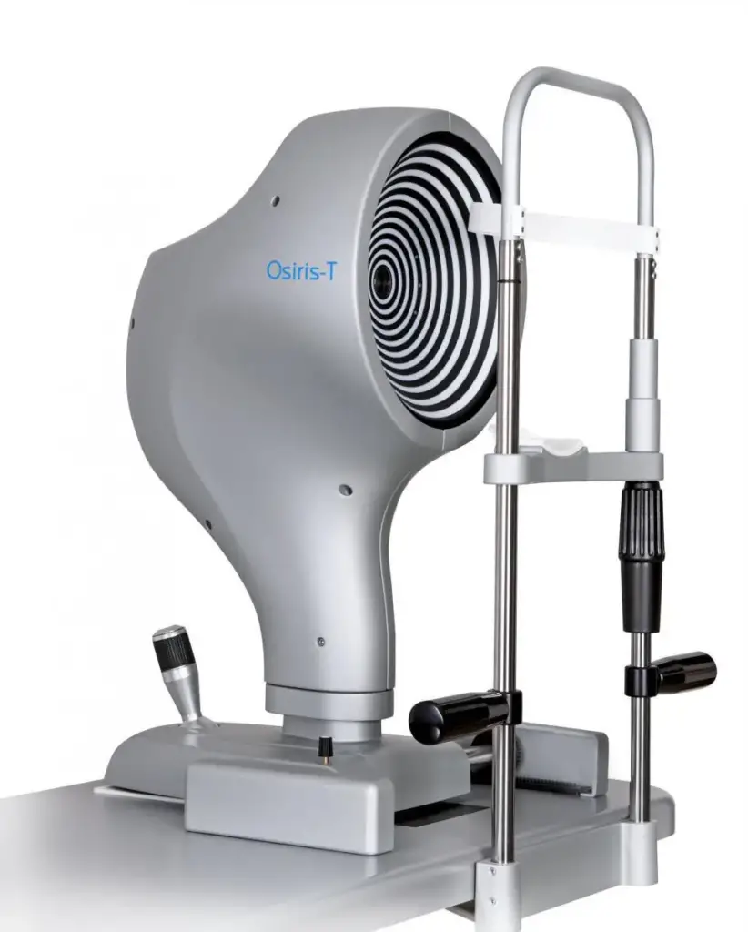
Corneal Topographer OSIRIS-T

Comprehensive Evaluation for Complex Ocular Aberrations
The OSIRIS-T corneal topographer, integrated with a total ocular aberrometer, offers essential information for accurately assessing patients with complex ocular aberrations, including both corneal and internal defects, in addition to traditional low-order defects.
Key Features:
Aberrometer:
- Unique Pyramidal Sensor Design: The OSIRIS-T aberrometer features a distinctive pyramidal sensor design, allowing for the measurement of aberrations with exceptional resolution—up to 45,000 points at the maximum pupil diameter—across a wide dynamic range.
- Real-time Wavefront Measurement: With a high frame rate of up to 33 images per second, the aberrometer can measure the ocular wavefront in real-time, capturing changes in power and aberrations as the patient accommodates.
- Phoenix Software Analysis: The Phoenix software provides an array of analysis options, including refractive error maps and visual simulations such as Point Spread Function (PSF), Modulation Transfer Function (MTF), and convolution with optotype. These tools aid clinicians in understanding and explaining patients’ visual problems effectively.
Topograph:
- Reflection Topography System: Utilizing a 22-ring Placido disk, the OSIRIS-T measures corneal morphology and refractive components through sagittal curvature, tangential curvature, elevation, and power maps.
- Synthesis Parameters: Consolidated synthesis parameters facilitate the straightforward diagnosis and follow-up of conditions like keratoconus, offering simplicity and intuitiveness in clinical assessments.
Toric Lens Assistant:
- Evaluation of Toric System Performance: By combining corneal topography data from CSO topographers with ocular aberration measurements, the OSIRIS-T enables clinicians to differentiate whether any astigmatic residue is attributable to lens rotation or incorrect calculation, enhancing the assessment of toric lens performance.
Applications:
- Diagnosis and Follow-up of Complex Ocular Aberrations: The OSIRIS-T provides indispensable information for evaluating patients with a wide range of ocular aberrations, aiding in accurate diagnosis and treatment planning.
- Evaluation of Toric System Performance: Clinicians can assess the performance of toric lens systems with confidence, distinguishing between astigmatic residue causes and ensuring optimal visual outcomes for patients.
Conclusion:
The Corneal Topographer OSIRIS-T offers clinicians a comprehensive solution for evaluating complex ocular aberrations, combining advanced aberrometry and topography capabilities. With its high-resolution measurements, real-time wavefront analysis, and intuitive software interface, the OSIRIS-T enhances diagnostic accuracy and facilitates personalized treatment strategies for improved patient care.
Follow us in Facebook
Discover more from An Eye Care Blog
Subscribe to get the latest posts sent to your email.

You must be logged in to post a comment.