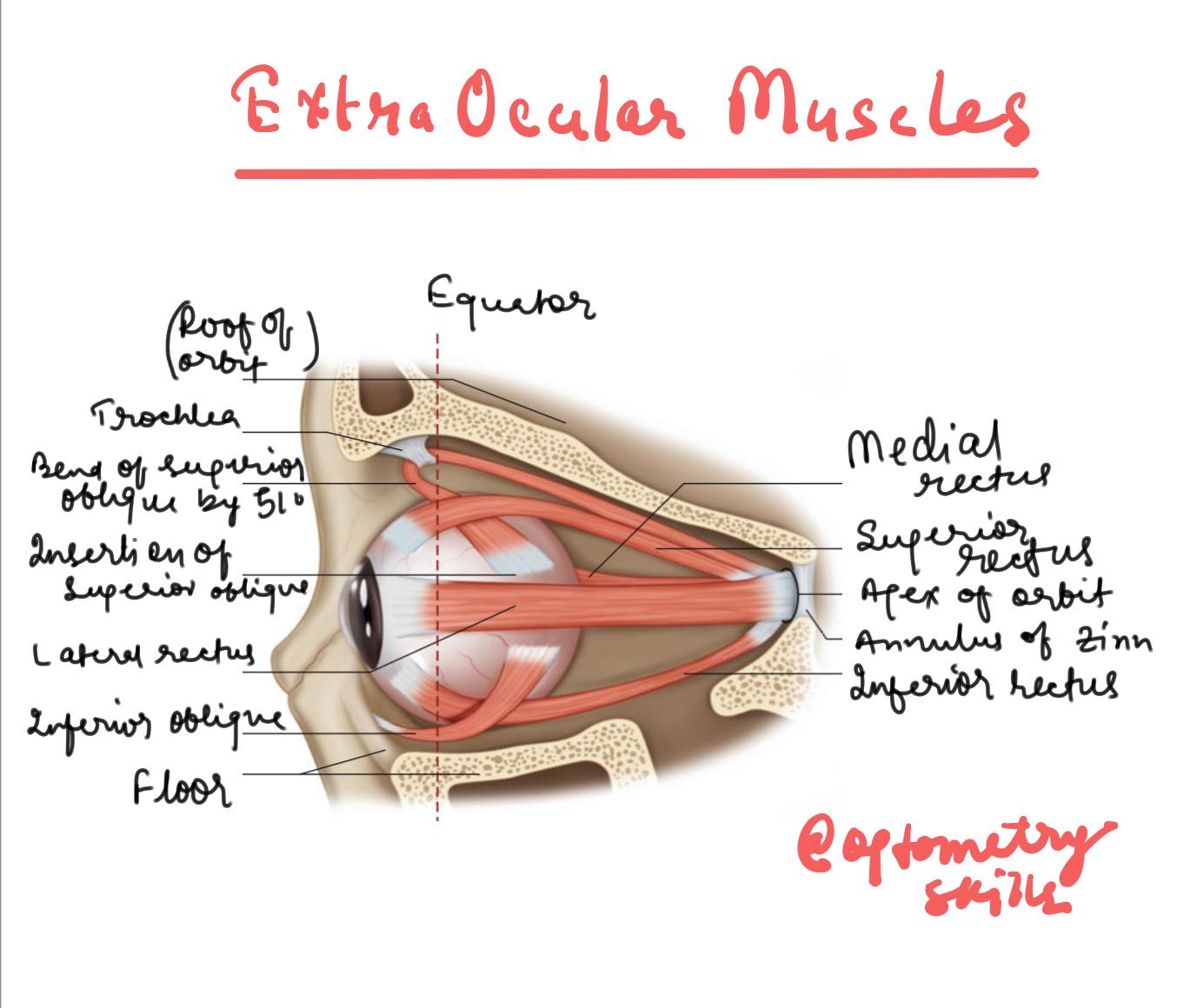
Diagram of 6 Extra Ocular Muscles

Understanding Extraocular Muscles
The human eye is a complex and fascinating organ, and its movement is controlled by a group of muscles known as the extraocular muscles. These muscles are responsible for the various movements of the eye, allowing us to look in different directions without moving our heads. Understanding the anatomy and function of these muscles is crucial, especially for professionals in optometry and ophthalmology.
Try MCQ Quiz on Extra ocular muscles
Anatomy of Extraocular Muscles
There are six extraocular muscles that control the movement of the eye, and they are categorized based on their location and the direction in which they move the eye. These muscles are:
- Medial Rectus: This muscle is located on the inner side of the eye and is responsible for moving the eye inward, towards the nose (adduction).
- Lateral Rectus: Positioned on the outer side of the eye, the lateral rectus muscle moves the eye outward, away from the nose (abduction).
- Superior Rectus: Situated on the top of the eye, this muscle elevates the eye, moving it upward. It also assists in turning the eye inward and rotates it slightly toward the nose.
- Inferior Rectus: Located below the eye, the inferior rectus muscle moves the eye downward. It also helps in adduction and rotates the eye slightly away from the nose.
- Superior Oblique: This muscle originates from the sphenoid bone and passes through a structure called the trochlea before inserting into the eye. The superior oblique muscle primarily moves the eye downward and outward, and it also rotates the top of the eye toward the nose (intorsion).
- Inferior Oblique: The inferior oblique muscle originates from the maxillary bone, located on the floor of the orbit. It moves the eye upward and outward and rotates the top of the eye away from the nose (extorsion).
The Role of the Extraocular Muscles
These muscles work in a coordinated manner to allow smooth and precise movements of the eye. The brain sends signals to the muscles to contract or relax, depending on the desired direction of gaze. For example, when you look to the right, the lateral rectus muscle of the right eye and the medial rectus muscle of the left eye contract simultaneously, allowing both eyes to move together.
Clinical Significance of understanding the location of 6 Extra Ocular Muscles
Understanding the function and anatomy of the extraocular muscles is vital in diagnosing and treating various eye conditions. Problems with these muscles can lead to issues such as strabismus (misalignment of the eyes), where the eyes do not properly align with each other. This can result in double vision or difficulty with depth perception.
Conclusion
The extraocular muscles play a crucial role in our ability to see the world around us. By allowing the eyes to move in different directions, they enable us to focus on objects, track moving targets, and maintain binocular vision. For optometrists, ophthalmologists, and other eye care professionals, a thorough knowledge of these muscles is essential for assessing and treating eye movement disorders.
Follow us in Facebook
Discover more from An Eye Care Blog
Subscribe to get the latest posts sent to your email.


You must be logged in to post a comment.