What do you See? Colour Vision test,Forms of Color Vision Deficiency
What is Colour Blindness?
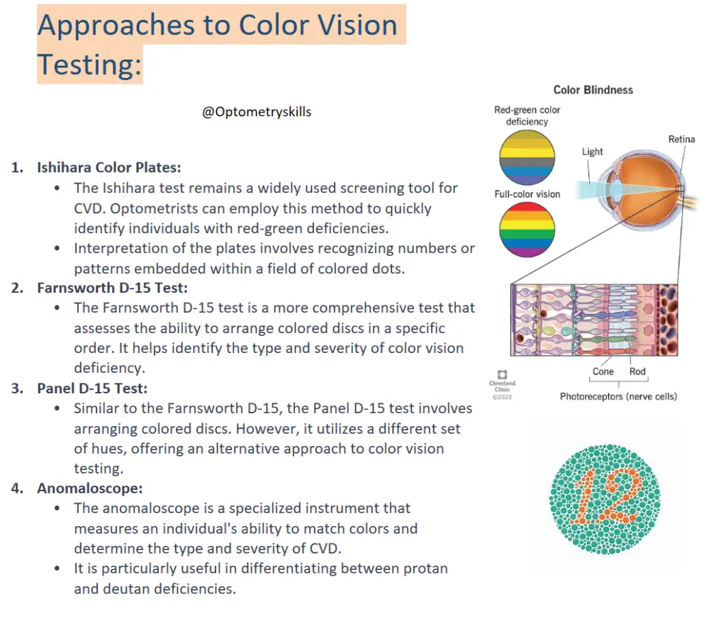
Color blindness is when you don’t see colors in the traditional way because some cones (nerve cells) in your eyes are missing or don’t work correctly. You may have trouble seeing the difference between certain colors or shades, or perceiving the brightness of colors. Color blindness is usually inherited through a genetic mutation.
Color vision is a complex process that involves the eyes and the brain working together to perceive the wide spectrum of colors present in the visual world. Color Vision Deficiency (CVD), commonly known as color blindness, occurs when there is a deficiency in the ability to perceive certain colors accurately. The three main types of cone cells in the retina responsible for color vision are sensitive to different wavelengths of light corresponding to the primary colors – red, green, and blue.
What are the symptoms of color blindness?
You might have a form of color blindness if you have trouble:
- Telling the difference between certain colors or shades.
- Seeing the brightness of certain colors.
But to recognize these symptoms, you need to know to look for them. Many people with color blindness don’t know to look for these differences because they’ve always seen colors in the same way. So, they don’t realize anything’s different about their color vision.
That’s why it’s important for children to have a comprehensive eye exam that includes colorblind testing before starting school. Many tests and other classroom materials rely on color to convey information or measure students’ learning. Children who see colors differently may struggle with these materials.

Ishihara Color Test (plates 2, 3, 6 and plate 10) is shown for a person with Protanopia blindness. (a) Original plates, (b) Images as seen by the color blindness (images can be retrieved from [15]).(ref)

Forms of Color Vision Deficiency:
- Protanopia and Protanomaly:
- Protanopia is a form of CVD where individuals have difficulty distinguishing between red and green colors. This results from the absence of functioning long-wavelength cones (L-cones).
- Protanomaly is a milder form, characterized by a reduced sensitivity to red light.
- Deuteranopia and Deuteranomaly:
- Deuteranopia is a CVD type where the green cones (M-cones) are missing, leading to challenges in differentiating between red and green hues.
- Deuteranomaly is a partial deficiency in M-cones, resulting in a reduced ability to perceive green light.
- Tritanopia and Tritanomaly:
- Tritanopia is a rare form of CVD where individuals struggle to distinguish between blue and yellow colors due to the absence of short-wavelength cones (S-cones).
- Tritanomaly is a milder form, involving a reduced sensitivity to blue light.
Primary Care Optometry Approaches to Color Vision Testing:
- Ishihara Color Plates:
- The Ishihara test remains a widely used screening tool for CVD. Optometrists can employ this method to quickly identify individuals with red-green deficiencies.
- Interpretation of the plates involves recognizing numbers or patterns embedded within a field of colored dots.
- Farnsworth D-15 Test:
- The Farnsworth D-15 test is a more comprehensive test that assesses the ability to arrange colored discs in a specific order. It helps identify the type and severity of color vision deficiency.
- Panel D-15 Test:
- Similar to the Farnsworth D-15, the Panel D-15 test involves arranging colored discs. However, it utilizes a different set of hues, offering an alternative approach to color vision testing.
- Anomaloscope:
- The anomaloscope is a specialized instrument that measures an individual’s ability to match colors and determine the type and severity of CVD.
- It is particularly useful in differentiating between protan and deutan deficiencies.
How common is Color Blindness?

Color blindness is more commonly observed in males compared to females, with the overall prevalence for males estimated to be around 8%, while for females, it is approximately 0.5%.
- Anomalous Trichromasy:
- Protanomaly, a form of red-green color deficiency, is found in about 1% of males and 0.01% of females.
- Deutanomaly, another red-green deficiency, is more common, affecting 5% of males and 0.4% of females.
- Tritanomaly, a blue-yellow deficiency, is considered rare and has negligible prevalence rates in both males and females.
- Dichromasy:
- Protanopia, characterized by the absence of red cones, has an estimated prevalence of 1% in males and 0.01% in females.
- Deuteranopia, the absence of green cones, affects approximately 1.5% of males and 0.01% of females.
- Tritanopia, the absence of blue cones, has a low prevalence of 0.008% in both males and females.
- Monochromasy:
- Rod monochromasy, cone monochromasy, and atypical monochromasy are all considered rare conditions. These conditions involve the absence or dysfunction of either rod or cone cells, leading to a more severe form of color blindness.
- These conditions are very rare, with no specific prevalence rates provided. However, the term “rare” indicates that they are infrequently encountered in the general population.
It’s important to note that the percentages provided are estimates and may vary in different populations. Additionally, the rarity of certain types of color vision deficiencies, such as tritanomaly, tritanopia, rod monochromasy, cone monochromasy, and atypical monochromasy, underscores the relative infrequency of these conditions compared to the more common red-green deficiencies (protanomaly, deutanomaly, protanopia, and deuteranopia).
Follow us in Facebook
Discover more from An Eye Care Blog
Subscribe to get the latest posts sent to your email.

You must be logged in to post a comment.