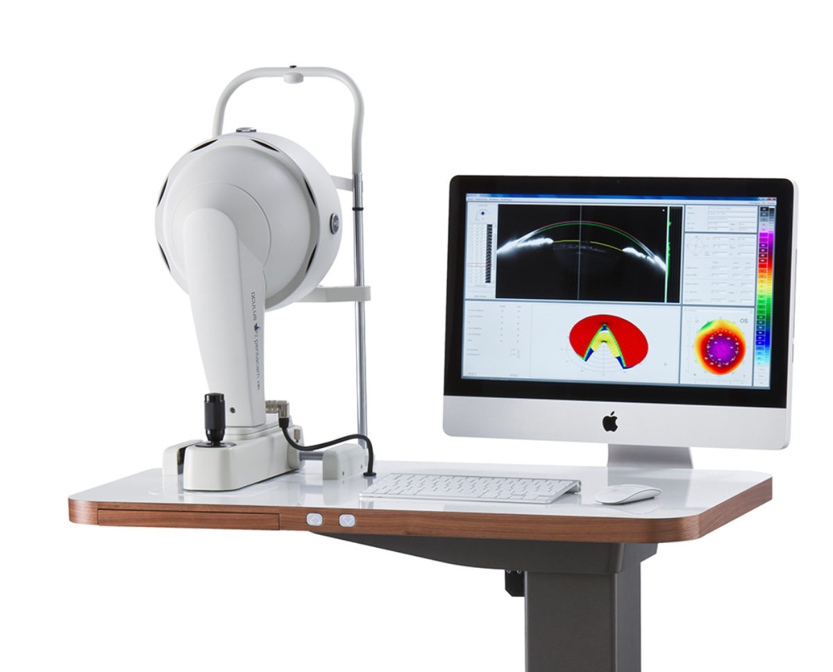
Pentacam eye screening
Pentacam is a non-invasive imaging technology that allows for comprehensive analysis of the cornea. It captures high-resolution images of the anterior segment of the eye, providing detailed information about the cornea’s curvature, thickness, and topography. In this article, we will explore the features and benefits of Pentacam and its applications in ophthalmology.
Pentacam topography is an advanced corneal imaging system that provides comprehensive analysis of the anterior segment of the eye. It was first introduced in the early 2000s and has since become a widely used device in ophthalmology clinics and research institutions.
The Pentacam offers non-invasive and high-resolution corneal measurements. It measures the surface of cornea and captures high-resolution images of the anterior segment of the eye, providing detailed information about the cornea’s curvature, thickness, and topography making it a reliable choice for diagnosing and monitoring various eye conditions. It can also help measure corneal astigmatism.

Terrien’s Marginal Corneal Degeneration
How does Pentacam works?

Pentacam is a corneal topography system that utilizes Scheimpflug imaging technology to capture high-quality images of the cornea. The system includes a rotating Scheimpflug camera that captures multiple images of the cornea from different angles. These images are then combined to create a 3D model of the cornea, allowing for detailed analysis of its structure and shape.

Pentacam eye
Corneal astigmatism can be measured using a Pentacam. Here’s how corneal astigmatism is typically measured with a Pentacam:
- Patient Preparation:
- Ensure the patient is properly positioned and comfortable.
- Instruct the patient to fixate on a target to keep their eye steady during the measurement.
- Instrument Setup:
- Calibrate the Pentacam according to the manufacturer’s instructions to ensure accurate measurements.
- Adjust the device’s settings as needed, such as selecting the appropriate mode (e.g., standard, high-resolution).
- Capture Corneal Data:
- The Pentacam uses a rotating Scheimpflug camera to capture multiple images of the cornea as it rotates around the eye. This allows it to create a 3D model of the cornea.
- Analysis:
- Once the data is captured, the Pentacam software analyzes the corneal shape and curvature, including any irregularities or astigmatism.
- The device measures various parameters, including anterior and posterior corneal curvature, keratometry readings, and corneal thickness.
- Astigmatism Measurement:
- Corneal astigmatism is typically reported as the difference in power between the steepest and flattest meridians of the cornea.
- The Pentacam provides astigmatism measurements in diopters (D) and can also display astigmatism in both the anterior and posterior corneal surfaces.
- Interpretation:
- Review the Pentacam-generated report to assess the degree and orientation of corneal astigmatism.
- This information is essential for preoperative planning in refractive surgeries like LASIK or toric IOL implantation, as well as for diagnosing and managing corneal conditions.
Pentacam Cost:
A Pentacam can cost you around $39,999.00. Also see 24 Eye examination Equipment for an Eye clinic.
Features of Pentacam
Pentacam offers a range of features that make it a valuable tool in ophthalmology. Some of its key features include:
- Corneal thickness measurement: Pentacam can measure corneal thickness with high accuracy, making it a valuable tool for diagnosing conditions such as keratoconus and glaucoma.
- Corneal topography: The system can create detailed maps of the cornea’s curvature, allowing for the detection of irregularities that may indicate conditions such as astigmatism or corneal ectasia.
- Anterior chamber analysis: Pentacam can also analyze the anterior chamber of the eye, providing information about the depth and volume of the chamber.
- Pupil analysis: The system can measure the size and shape of the pupil, providing valuable information for procedures such as LASIK surgery.
Applications of Pentacam
Pentacam has a range of applications in ophthalmology, including:
- Diagnosis of corneal conditions: Pentacam can be used to diagnose a range of corneal conditions, including keratoconus, corneal dystrophies, and corneal ectasia.
- Pre-operative planning for refractive surgery: Pentacam can provide detailed information about the cornea’s curvature and thickness, allowing for precise pre-operative planning for procedures such as LASIK surgery.
- Monitoring of corneal changes: Pentacam can be used to monitor changes in the cornea over time, allowing for early detection of conditions such as keratoconus and glaucoma.
- Evaluation of contact lens fit: Pentacam can be used to evaluate the fit of contact lenses, ensuring that they are comfortable and do not damage the cornea.
Benefits of Pentacam
Pentacam offers a range of benefits over traditional corneal imaging technologies, including:
- Non-invasive: Pentacam is a non-invasive imaging technology that does not require any contact with the eye, making it comfortable and safe for patients.
- High accuracy: Pentacam can capture detailed images of the cornea with high accuracy, allowing for precise diagnosis and treatment planning.
- Comprehensive corneal analysis: Pentacam captures the structures in the entire cornea and provides information about corneal thickness in multiple areas of the cornea.
- Quick and easy: Pentacam imaging is quick and easy, taking only a few minutes to complete.
- Comprehensive analysis: Pentacam provides a comprehensive analysis of the cornea and anterior chamber, allowing for the detection of a range of conditions and abnormalities.
Comparison with Other Anterior Eye Segment Tomography Devices:
The Pentacam has several advantages over other anterior eye segment tomography devices. Compared to ultrasound-type techniques, Pentacam offers higher resolution, non-contact measurement, and does not require anesthesia. Compared to optical coherence tomography (OCT) techniques, Pentacam provides more comprehensive corneal examination and offers more significant analysis.
Clinical Applications of Pentacam:
The Pentacam has various clinical applications, including measuring corneal thickness as a screening tool for LASIK surgery, detecting early signs of corneal diseases, and evaluating the effectiveness of the treatment. It is also used for pre-operative assessment of cataract and refractive surgery, diagnosing and monitoring keratoconus, measuring IOP, and diagnosing and treating dry eyes.
Pentacam models
Pentacam®: Fast Screening Report, Topography and Elevation Maps of the Anterior and Posterior Corneal Surface, Pachymetry Maps (absolute and relative), Scheimpflug Image Overview, 3D Anterior chamber analysis, Anterior segment tomography, Topographical KC Classification (TKC), Belin ABCD KC Staging, Belin ABCD Progression Display, Iris Image and HWTW, 4 Maps Refractive, Compare 2 Exams, Belin/Ambrósio Display, Contact Lens Fitting.
Pentacam®
The Pentacam® provides you with an overall view of the anterior eye segment in a matter of seconds. Measurements are performed by auto-release and are accompanied by a quality test, thus guaranteed to be fast, reproducible and delegable.
The Pentacam® comes with an extensive basic software package which can be extended according to your needs to include further optional software packages and modules.
- Fast Screening Report
- Contact Lens Fitting
- Belin/Ambrósio Display
- Topographical KC Classification (TKC)
- Tomographical KC Classification
- Belin ABCD KC Staging
- Belin ABCD Progression Display
- Qualitative assessment of the cornea
- Topography and Elevation Maps of the Anterior and Posterior Corneal Surface
- overall pachymetry
- Anterior chamber angle screening:
- pachymetry-based IOP correction
- chamber angle and chamber volume
- Elevation data
- Comparative displays for follow-up
- Comparison and superimposition of Scheimpflug images
Pentacam® HR: Includes all the features of the Pentacam® model plus additional optional software modules such as CSP Report, Refractive Package, Cataract Package, Holladay Report and Holladay EKR65 Detail Report, PNS and 3D Cataract Analysis, Corneal Optical Densitometry, IOL Calculator and 3D pIOL Simulation and Aging Prediction.
With its brighter and optimized optics the Pentacam® HR offers you brilliant image quality.The resolution of its images is five times that of the Pentacam® models, enabling the Pentacam® HR to deliver impressive representations of IOLs and phakic IOLs.
The Pentacam® HR comes with an extensive basic software package which can be extended according to your needs to include further optional software packages and modules.
Even the basic software offers a vast range of functions.
- Fast Screening Report
- Contact Lens Fitting
- Belin/Ambrósio Display
- Topographical KC Classification (TKC)
- Tomographical KC Classification
- Belin ABCD KC Staging
- Belin ABCD Progression Display
- Qualitative assessment of the cornea
- Topography and Elevation Maps of the Anterior and Posterior Corneal Surface
- overall pachymetry
- Anterior chamber angle screening:
- pachymetry-based IOP correction
- chamber angle and chamber volume
- Elevation data
- Comparative displays for follow-up
- Comparison and superimposition of Scheimpflug images
Pentacam® AXL: Includes all the features of the Pentacam® HR model.
Within a mere 2 seconds the Pentacam® AXL supplies you with precise diagnostic data on the entire anterior eye segment. In addition to anterior segment tomography the Pentacam® AXL has integrated axial length measurement, a feature which allows you to make accurate IOL calculations. Automatic measurement activation with quality test guarantees fast, reproducible and delegable measurements.
The Pentacam® AXL comes with an extensive basic software package which can be extended according to your needs to include further optional software packages and modules.

The Pentacam® AXL determines the axial length of the eye as well as the data of the anterior eye segment, from the anterior corneal surface to the posterior surface of the crystalline lens, all in a single measurement. In doing so it makes combined use of the time-tested Pentacam® technology and an equally proven interferometry-based biometry technique. The Pentacam® AXL is the right choice for those seeking to perform anterior-segment diagnostics and IOL calculations using one and the same instrument.
Even the basic software offers a vast range of functions.
- IOL Calculator for
- both treated and untreated eyes
- spherical, aspherical multifocal and toric IOL calculation
- toric IOL calculation based on total corneal refractive power
- Fast Screening Report
- Belin/Ambrósio Display
- Topographical KC Classification (TKC)
- Tomographical KC Classification
- Belin ABCD KC Staging
- Belin ABCD Progression Display
- Qualitative assessment of the cornea
- Topography and Elevation Maps of the Anterior and Posterior Corneal Surface
- overall pachymetry
- Corneal Optical Densitometry
- Anterior chamber angle screening
- pachymetry-based IOP correction
- chamber angle and chamber volume
- Elevation data
- PNS and 3D Cataract Analysis
- Premium IOL selection in 4 steps
- Comparative displays for follow-up
- Comparison and superimposition of Scheimpflug images
- Contact Lens Fitting
Pentacam® AXL Wave: Includes all the features of the Pentacam® AXL model.
The new Pentacam® AXL Wave provides the unique combination of:
- Scheimpflug based Tomography
- Optical Biometry
- Wavefront Aberrometry of the entire eye
- Objective Refraction
- Retroillumination
Pre- op help it can provide to the surgeon
- One single intuitive scanning process
- Comprehensive screening for possible diseases and abnormalities
- Optical corneal and lens evaluation
- Clear patient education
- Optimal premium IOL selection based on real measurements
- Most up to date IOL power calculation for all corneal shapes
OPtimal post op assessment
- IOL inclination and centration
- Objective refraction
- Quality assessment and improvement
Sirius Pentacam
the “Sirius Scheimpflug-Placido Topographer.” It is produced by the Swiss company CSO (Costruzione Strumenti Oftalmici), which specializes in manufacturing ophthalmic equipment.
The Sirius Pentacam is an advanced ophthalmic imaging system used primarily in eye care and ophthalmology practices. It combines two technologies: Scheimpflug imaging and Placido disc topography. Here’s a brief explanation of each technology:
- Scheimpflug Imaging: Scheimpflug imaging is a technique used to capture images of the anterior segment of the eye. It works by projecting a slit of light onto the eye at an angle and then capturing the image of the illuminated section. The imaging technique is named after the Austrian physicist Theodor Scheimpflug, who first described it in 1904. The Sirius Pentacam uses this technology to obtain high-resolution 3D images of the cornea, anterior chamber, and lens.
- Placido Disc Topography: Placido disc topography is a method for measuring the shape and curvature of the cornea. It involves projecting a series of concentric rings onto the cornea and observing their reflection. By analyzing the distortion of the reflected rings, the device can calculate the corneal curvature and create a detailed corneal topography map.
The combination of Scheimpflug imaging and Placido disc topography in the Sirius Pentacam allows eye care professionals to obtain comprehensive and precise information about the cornea and anterior segment of the eye. It is used for various purposes, including:
- Assessing the corneal shape and curvature for refractive surgery planning (e.g., LASIK or PRK).
- Diagnosing and monitoring corneal conditions, such as keratoconus or pterygium.
- Evaluating the anterior chamber and assessing the angle for glaucoma screening.
- Determining the suitability of patients for intraocular lens (IOL) implantation before cataract surgery.
- Monitoring changes in corneal thickness and shape over time.
The Sirius Pentacam is considered a valuable tool for comprehensive eye examinations and aids in the accurate diagnosis and management of various eye conditions. It is commonly used by ophthalmologists and eye care professionals to provide optimal care for their patients.
oculus pentacam

Since its introduction in 2002, the OCULUS Pentacam® has proven itself indispensable and has come to represent the “Gold Standard” worldwide.
OCULUS Pentacam HR offers you excellent image quality. The resolution of its images is five times that of the Pentacam Basic or even Classic models, allowing the Pentacam HR to deliver efficient representations of IOLs and IOLs.
Within just two seconds the Pentacam® supplies you with precise diagnostic data on the entire anterior eye segment. The degree of corneal or crystalline lens density is made visible by the light scattering properties of the crystalline lens and is automatically quantified by the software.
Measurement of the anterior and posterior corneal surfaces supplies the total refractive power as well as the thickness of the cornea over its entire area. The data on the posterior surface provide optimal assistance in the early detection of ectatic changes.
The rotating scan supplies a large number of data points in the center of the cornea. A supplementary pupil camera captures eye movements during the examination for subsequent automatic correction of measured data.
Also read: Studies on Pentacam
Follow us in Facebook


