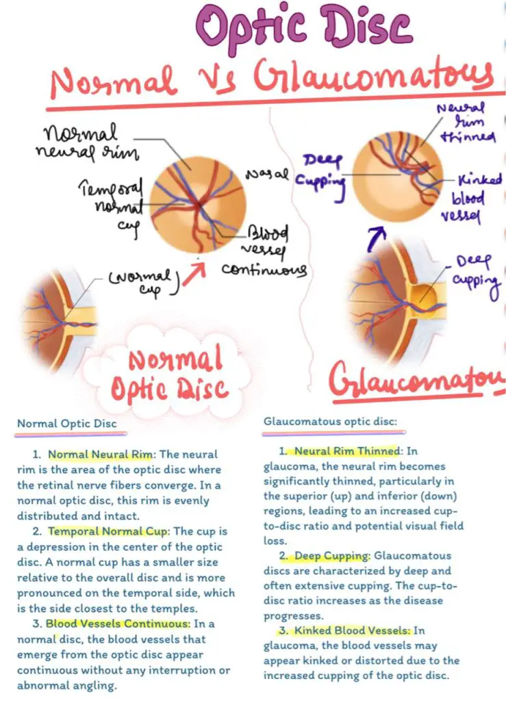
Difference in Optic Disc of Normal and glaucomatous Disc

Understanding the Difference in Optic Disc of Normal and glaucomatous Disc
The optic disc, also known as the optic nerve head, is a crucial structure in the eye that can reveal much about a person’s ocular health. Differentiating between a normal optic disc and one affected by glaucoma is essential for the diagnosis and management of the disease.
Normal Optic Disc
A healthy optic disc is typically characterized by a small, central depression called the “cup,” surrounded by a reddish neural tissue, which transmits visual information to the brain. The cup-to-disc ratio (the size of the cup relative to the entire optic disc) is usually small, reflecting the presence of a thick layer of nerve fibers. The appearance of the optic disc can vary, but the overall structure should show no signs of damage, with a consistent, symmetrical pattern in both eyes.
Glaucomatous Optic Disc
In glaucoma, the optic nerve becomes progressively damaged, leading to a characteristic change in the optic disc’s appearance. The most notable feature is increased cupping, where the cup-to-disc ratio enlarges due to the loss of neural tissue. This results in a larger, more pronounced cup and a thinner neural rim. As glaucoma advances, the optic disc may show notching or thinning, especially at the superior and inferior poles. Additionally, there might be visible disc hemorrhages and areas of pallor, indicating advanced nerve fiber loss.
One of the significant challenges in diagnosing glaucoma is distinguishing these changes from non-glaucomatous optic neuropathies, which can also cause cupping but are usually associated with other symptoms like acute vision loss or color vision defects. Non-glaucomatous cupping tends to be more diffuse or located at the temporal side of the optic disc, unlike the localized notching seen in glaucomatous damage.

Glaucomatous Optic Disc
In glaucoma, a disease that damages the optic nerve, the appearance of the optic disc changes significantly. These changes include:
- Neural Rim Thinned: In a glaucomatous optic disc, the neural rim becomes significantly thinned, particularly in the superior (upper) and inferior (lower) regions. This thinning leads to an increased cup-to-disc ratio, which is a key indicator of glaucoma. As the disease progresses, the thinning of the neural rim may result in visual field loss.
- Deep Cupping: Glaucoma is characterized by deep and often extensive cupping of the optic disc. The cup-to-disc ratio increases as the disease progresses, reflecting the loss of nerve fibers and the corresponding deepening of the optic cup.
- Kinked Blood Vessels: In glaucoma, the blood vessels on the optic disc may appear kinked or distorted due to the increased cupping. This kinking is a result of the structural changes in the optic disc caused by the disease.
Importance of Early Detection
Regular eye exams and early detection of glaucomatous changes are vital in preventing irreversible vision loss. While normal optic discs maintain their structure over time, glaucomatous discs demonstrate a progressive enlargement of the cup and loss of neural tissue, making early diagnosis critical for effective treatment.
Follow us in Facebook
Discover more from An Eye Care Blog
Subscribe to get the latest posts sent to your email.

You must be logged in to post a comment.