What is Meibography ?
Meiobography, also known as meiobian gland dysfunction or MGD, is a common eye condition that affects millions of people worldwide. Meibomian gland dysfunction (MGD) is now considered the leading cause of dry eye.
Meiobian glands are small glands located on the eyelids that produce the oily layer of the tear film. This oily layer helps to keep the water in tears from evaporating too quickly, which can lead to dry eye syndrome. When these glands become blocked or dysfunctional, it can result in meiobian gland dysfunction or MGD.
MGD is a chronic condition that can cause discomfort, irritation, and even vision problems. It is typically characterized by symptoms such as dry eyes, redness, itching, and a burning sensation in the eyes. Left untreated, MGD can lead to more severe conditions such as corneal ulcers, infections, and even vision loss.
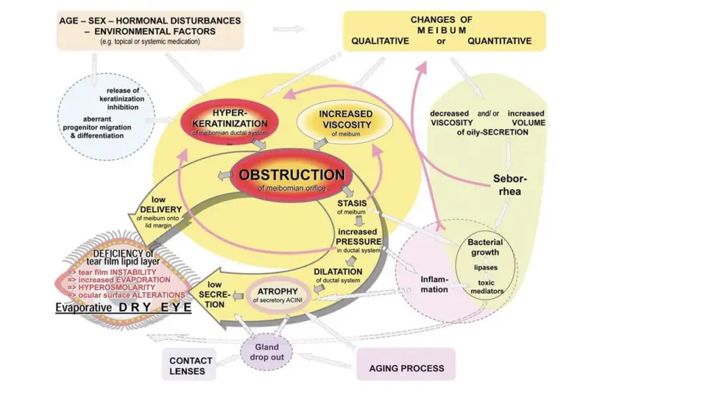
Pathways involved in the pathophysiology of meibomian gland dysfunction (MGD) proposed by the 2011 International Workshop on Meibomian Gland Dysfunction:
Credit: Baudouin C, Messmer EM, Aragona P, et al. Revisiting the vicious circle of dry eye disease: a focus on the pathophysiology of meibomian gland dysfunction. Br J Ophthalmol 2016;100:300-6.
Meibomian gland dysfunction (MGD) cause and treatment
Meibomian gland dysfunction (MGD) is a condition where the meibomian glands within the eyelids become blocked or dysfunctional. This can lead to a decrease in the production of the lipid-rich substance that helps to protect the tear film. As a result, the tear film evaporates quickly, leading to dry eyes and other symptoms.
The causes of MGD can vary and may include factors such as age, hormonal changes, environmental factors, and certain medical conditions. Symptoms of MGD can be mild or severe and may include dry eyes, burning or stinging sensations in the eyes, gritty or sandy sensations in the eyes, and blurred vision.
Meibography Techniques and Technologies

Meibography is a non-invasive imaging technique that is used to visualize the meibomian glands within the eyelids. This technique can help clinicians to assess the health of the meibomian glands and to diagnose MGD.
There are several meibography techniques and technologies available today. One of the most common techniques is infrared meibography, which uses infrared light to visualize the meibomian glands. Other techniques are-
- LipiView Interferometer: This instrument uses advanced technology to capture high-resolution images and videos of the meiobian glands. It allows eye doctors to evaluate the function and structure of the glands and identify any blockages or abnormalities.
- Meibomian Gland Evaluator: This handheld device is used to apply pressure to the meiobian glands and assess their function. It can help identify any issues with gland blockages or dysfunction.
- TearLab Osmolarity System: This instrument measures the osmolarity, or salt concentration, of tears. Elevated osmolarity levels are often associated with dry eye syndrome and MGD.
- Infrared Meibography: This technology uses infrared light to capture images of the meiobian glands. It can help identify any blockages or structural abnormalities within the glands.
How to Assess Meibomian Gland Function Using Diagnostic Expression of the Glands
In addition to meibography, there are other methods for assessing meibomian gland function. One such method is diagnostic expression of the glands, where pressure is applied to the eyelids to express the meibomian gland secretions. The quantity and quality of the secretions can then be evaluated.
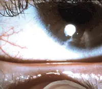
A normal healthy eyelid. Upon gentle pressure over the glands, the gland orifices release clear liquid oil.
Another method is tear film lipid layer evaluation, which assesses the thickness and quality of the lipid layer that covers the tear film. This can be done using specialized instruments such as the LipiView® interferometer.
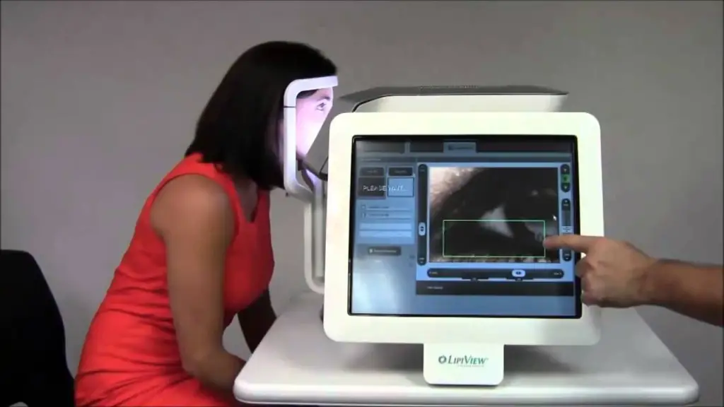
Capturing and Analyzing Meibography Images: How to Capture and Interpret Meibography Images?
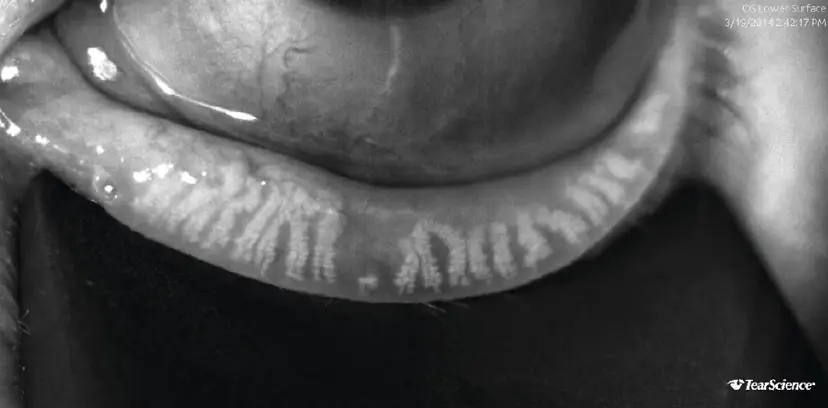
Lipiscan showing meibomian gland atrophy
Capturing meibography images requires specialized equipment and training. Typically, a meibography device is used to capture images of the meibomian glands. The patient’s eyelids are gently pulled down, and the device is placed against the eyelid. Images are then captured using infrared light or another imaging technique.
Once meibography images are captured, they can be analyzed to assess the health of the meibomian glands. Clinicians can use these images to evaluate the density and morphology of the glands, as well as the level of gland dropout. This information can be used to diagnose MGD and to develop a treatment plan.
Treatment
There is treatment available for Meibography. You should see an ophthalmologist and get appropriate treatment. At home you can do warm compress and remember not to rub the eyes after warm compress and drink plenty of fluid and keep hydrated.
References:
- Nichols, Jason J., et al. “The international workshop on meibomian gland dysfunction: executive summary.” Investigative Ophthalmology & Visual Science 52.4 (2011): 1922-1929.
- Blackie, Caroline A., et al. “Nonobvious obstructive meibomian gland dysfunction.” Cornea 28.12 (2009): 1346-1353.
- Pult, Heiko, and James S. Wolffsohn. “The ocular surface in keratoconjunctivitis sicca.” Contact Lens and Anterior Eye 33.5 (2010): 157-165.
Follow us at Optometryskills for daily updates
Discover more from An Eye Care Blog
Subscribe to get the latest posts sent to your email.


You must be logged in to post a comment.