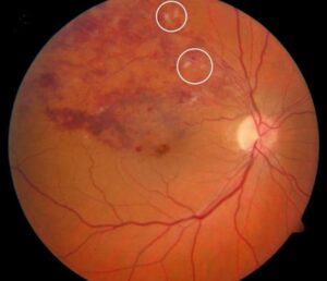
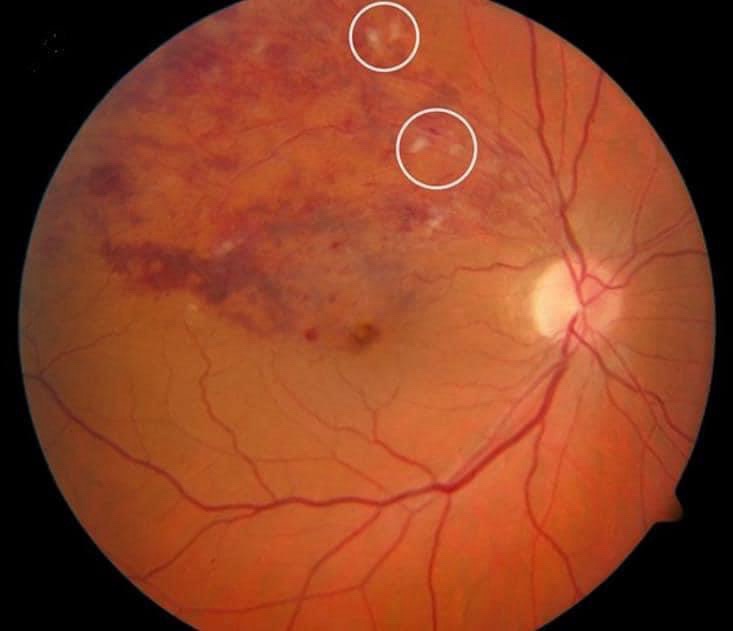
Diagnosis challenge- Is this Cotton wool spot or exudate?
What are the circled area in the picture?

We had a diagnosis challenge in out facebook page. Some said it is cotton wool spot and some said it is exudate. So how do we differentiate between the two and what is the correct answer?
The circled areas in the image are likely cotton wool spots.
Here’s why:
- Cotton wool spots are soft white patches on the retina caused by ischemia (lack of blood supply) to the nerve fiber layer. They result from occlusion of small retinal arterioles, which leads to axoplasmic material build-up within the retinal nerve fibers.
- Exudates, on the other hand, are yellowish deposits made of lipids and proteins that leak from damaged blood vessels. These are typically found in a ring-like pattern around areas of retinal swelling, especially in conditions like diabetic retinopathy.
Since the circled spots appear whiter and are isolated, rather than the more yellowish appearance of exudates, it’s more consistent with cotton wool spots. Cotton wool spots are commonly seen in retinal vein occlusions due to capillary non-perfusion and the resulting nerve fiber damage.
The fundus image is that of BRVO
Branch Retinal Vein Occlusion (BRVO) is a common retinal vascular disease that occurs when one of the smaller veins in the retina becomes blocked, leading to impaired blood flow and swelling in the area. This condition affects the part of the retina that is served by the blocked vein, and is usually caused by underlying issues like hypertension, atherosclerosis, or diabetes.
Key Points
- Risk factors include age, high blood pressure, and cardiovascular diseases.
Symptoms:
- Sudden, painless loss of vision in the affected eye, usually in part of the visual field.
- Blurred or distorted vision.
- In some cases, the condition may be asymptomatic and detected during routine eye exams.
Diagnosis:
- Fundus Photography: Shows retinal hemorrhages (bleeding), cotton wool spots, and dilated veins
- Optical Coherence Tomography (OCT): Useful for assessing retinal thickness and macular edema.
- Fluorescein Angiography: Can help assess the blood flow and leakage in the retina.
Management:
- Regular monitoring to assess for complications like macular edema.
- Laser Treatment
- Anti-VEGF Injections: To reduce macular edema and improve vision.
- Steroid Injections: Can also be used to control inflammation and swelling.
Prognosis:
Many patients experience some visual improvement after treatment, but the degree of recovery can vary depending on the severity of the occlusion and any underlying health conditions.
Discover more from An Eye Care Blog
Subscribe to get the latest posts sent to your email.


You must be logged in to post a comment.