Vitreous Cyst: A Rare and Fascinating Ocular Finding
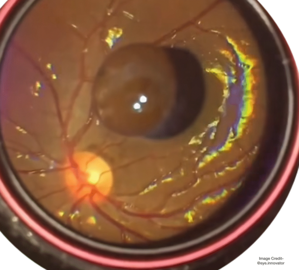
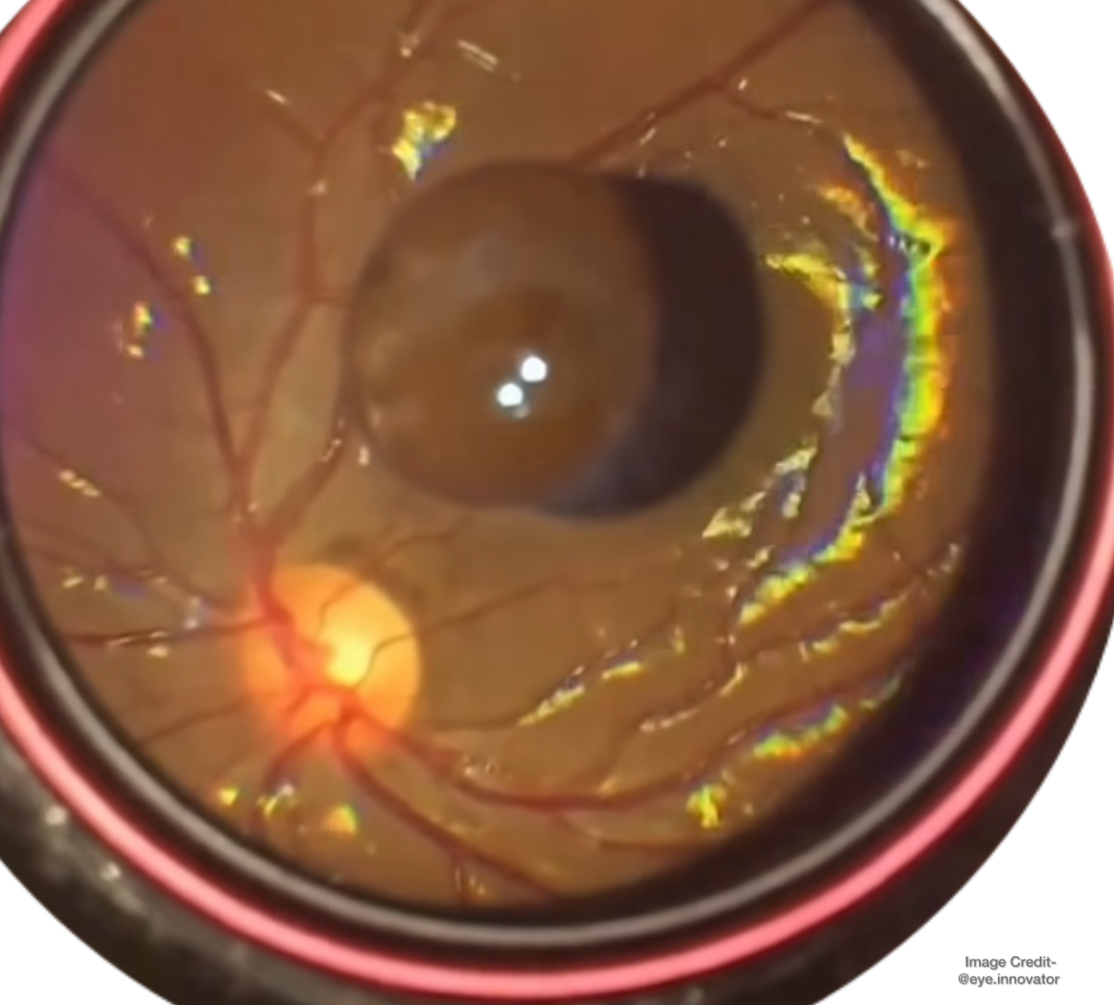
A 20-year-old female presented with complaints of floaters in her right eye. On fundus examination, a smooth, pigmented, mobile cystic body was observed near the optic disc — a classic presentation of a vitreous cyst.
What makes this case interesting is the absence of any history of trauma or ocular inflammation. Vitreous cysts are uncommon intraocular findings that may be congenital or acquired, and most often, they remain benign and non-progressive.

Understanding Vitreous Cysts
Vitreous cysts are rare, free-floating or attached cystic structures within the vitreous cavity. They can vary in color, transparency, and mobility, depending on their origin and content.
Congenital vitreous cysts are believed to originate from choristomas of the primary hyaloid system, derived from the iris or ciliary pigment epithelium. Histologically, they contain immature melanosomes, which explains their pigmented appearance.
Acquired cysts, on the other hand, may develop secondary to ocular trauma, parasitic infections (such as cysticercosis), or inflammation. However, in this case, none of these were present, suggesting a congenital origin.
Clinical Presentation
Patients often present with:
Floaters or visual disturbances Normal visual acuity in most cases A mobile cyst visible on fundus examination
The cyst can be either free-floating or attached to the optic disc, ciliary body, or retina. Fundus photography and ultrasonography help confirm the diagnosis and rule out other intraocular lesions.
Management Approach
The management of vitreous cysts depends on symptoms and patient comfort.
Asymptomatic cases: Observation and periodic follow-up are usually sufficient, as most cysts remain stable without intervention.
Symptomatic cases: If the cyst significantly interferes with vision, two main options exist: Laser cystotomy (Argon or Nd:YAG) — to rupture or collapse the cyst. Pars plana vitrectomy (PPV) with cyst excision — for definitive removal in persistent or bothersome cases.
Prognosis
The overall prognosis for vitreous cysts is excellent. Most remain unchanged for years and do not lead to visual loss or other complications. Regular monitoring ensures early detection of any changes in size or mobility.
Credits: @eye.innovator
Note
“The overview above is consistent with current literature: vitreous cysts are rare, often congenital, and usually benign. However, the precise origin is not always proven and cases should be evaluated with multimodal imaging to exclude infectious or neoplastic causes. Management ranges from observation for asymptomatic patients to laser photocystotomy (Argon or Nd:YAG) or pars plana vitrectomy for persistent or visually significant lesions. Both laser and surgical options have benefits and risks and should be chosen case-by-case.
You can read more on this case here:
EyeWiki — Vitreous Cysts (overview). Primary vitreous cysts — BMC Ophthalmology (2024) — multimodal imaging paper.
Primary vitreous cysts — Robben et al., case reports with histopathology & imaging (2020, PMC).
Idiopathic pigmented vitreous cyst — BMC Ophthalmology / case series (electron microscopy supporting pigment epithelial origin).
JAMA Ophthalmology — “Idiopathic Pigmented Vitreous Cyst” (case and management discussion).
Discover more from An Eye Care Blog
Subscribe to get the latest posts sent to your email.


You must be logged in to post a comment.